
Page 16 - v11i4
P. 16
International Journal of Bioprinting 3D-printed scaffolds for osteochondral defect
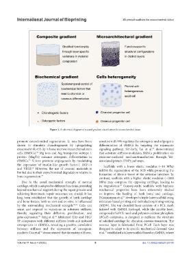
Figure 1. A schematic diagram of natural gradient classification for osteochondral tissue.
promote osteochondral regeneration. Li ions have been sensitive miR-99b regulates the osteogenic and adipogenic
shown to stimulate chondrogenesis by upregulating differentiation of BMSCs by targeting the rapamycin
exosomal miR-455-3p in bone marrow mesenchymal stem signaling pathway. Similarly, Lai et al. demonstrated
83
cells (BMSCs). Mg ions and Mg transporter subtype 1 that substrate stiffness modulates BMSCs proliferation via
74
protein (MagT1) enhance osteogenic differentiation in exosome-mediated mechanotransduction through Yes-
rBMSCs. Si ions promote angiogenesis by modulating associated protein (YAP) pathways.
75
the expression of insulin-like growth factor-1 (IGF-1) Scaffolds with a lower elastic modulus (~10 MPa)
and VEGF. However, the use of ceramic materials is inhibit the regeneration of the SCB while promoting the
76
limited due to their asynchronized degradation relative to formation of fibrous tissue at the articular interface. In
bone regeneration. 77
contrast, scaffolds with a higher elastic modulus (~1000
Due to the zonal mechanical strength of normal MPa) may compress the opposing cartilage, leading to
84
cartilage, which is adapted to different functions, providing its degradation. Consequently, scaffolds with biphasic
layered mechanical support during the repair process and mechanical properties have been extensively studied
achieving biomimetic repair outcomes are crucial. It has to improve the healing of both bone and cartilage.
long been established that the growth of both cartilage Diloksumpan et al. developed a triple-layer scaffold using
45
and bone tissues, both in vivo and in vitro, is influenced extrusion-based printing and melt electrospinning writing
by the surrounding mechanical strength. 78,79 Cells can (MEW). The top chondral layer consists of a PCL mesh
sense and respond to variations in substrate stiffness, infused with GelMA hydrogel, while the middle layer,
thereby regulating their diffusion, proliferation, and composed of a PCL mesh and polymer-calcium phosphate
gene expression. Yang et al. fabricated HAp and PEG/ (PCaP) composite, is designed to replicate the structure
81
80
SF composites with different stiffness levels and induced of calcified cartilage for chondral-osseous transition. The
osteogenesis in rBMSCs, revealing a positive correlation osseous layer is fabricated from PCaP. Each layer was
between stiffness and the expression of osteogenic designed to adapt to its specific mechanical demand. Gao
markers. Cao et al. demonstrated that the matrix stiffness- et al. established a bilayer scaffold based on GelMA, where
82
47
Volume 11 Issue 4 (2025) 8 doi: 10.36922/IJB025120100