
Page 300 - v11i4
P. 300
International Journal of Bioprinting Dual tuning of 3D-printed SilMA hydrogel
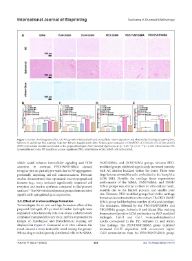
Figure 7. In vitro chondrogenesis effect. (A) The growth of internal cells and extracellular matrix deposition was observed by histological staining (HE,
Safranin O, and Alcian Blue staining). Scale bar: 100 µm; magnification: 200×. Relative gene expression of (B) MKI67, (C) COL2A1, (D) ACAN, and (E)
SOX9 of articulator chondrocytes loaded in the prepared hydrogels. Note: Statistical significance at *p < 0.05, **p < 0.01, ***p < 0.001. Abbreviations: HE,
hematoxylin and eosin; NF, nanofibers; ns, non-significant; PEO, poly(ethylene oxide); SilMA, silk methacryloyl.
which could enhance intercellular signaling and ECM 1%NF/SilMA, and 2%NF/SilMA groups, whereas PEO-
secretion. In contrast, PEO/2%NF/SilMA showed modified groups exhibited significantly increased porosity
irregular pits on partial pore walls due to NF aggregation, with AC clusters localized within the pores. There were
potentially impeding cell-cell communication. Previous large lacunae around the cells, embedded in the basophilic
studies demonstrated that optimized microtopographical ECM (HE). Notably, the cartilage tissue regeneration
features (e.g., wavy surfaces) significantly improved cell performance of the SilMA, 1%NF/SilMA, and 2%NF/
retention and matrix synthesis compared to flat/grooved SilMA groups was similar to their in vitro culture result,
surfaces. The NF-reinforced porous groups demonstrated possibly due to the limited porosity and smaller pore
51
significantly upregulated gene expression. size. However, PEO-modified groups had visible cartilage
formation in contrast with in vitro culture. The PEO/1%NF/
3.5. Effect of in vivo cartilage formation SilMA group had the highest number of cells and cartilage-
To investigate the in vivo cartilage formation effect of the like structures, followed by the PEO/2%NF/SilMA and
prepared hydrogels, 3D-printed AC-laden hydrogels were PEO/SilMA groups. Safranin O and Alcian Blue staining
implanted subcutaneously into non-obese diabetic/severe demonstrated positive ECM production in PEO-modified
combined immunodeficiency mice, and the representative hydrogels. Col-II and Col-I immunohistochemical
images of histological and histochemical staining are results correspond to the HE, Safranin O, and Alcian
displayed in Figure 8. Compared to in vitro cultures, the Blue findings. The PEO/1%NF/SilMA group exhibited
result showed a more noticeable trend among the groups. increased Col-II deposition with concurrent higher
HE staining revealed sparsely distributed cells in the SilMA, Col-I accumulation than the PEO/2%NF/SilMA group.
Volume 11 Issue 4 (2025) 292 doi: 10.36922/IJB025140118