
Page 438 - v11i4
P. 438
International Journal of Bioprinting Osteocytic Wnt7b-PKCδ against microgravity
2.6. Alkaline phosphatase staining 2.8. Mineralization assay
Alkaline phosphatase (AP) staining was performed as Mineralized matrix deposition was assessed by ARS
23
described previously. Cells were washed with 1× PBS and staining, as previously described. Osteocytes and ST2
23
fixed with 3.7% formaldehyde (Chongqing Chuandong cells were seeded in 24-well plates or PCI3D modules
Chemical Group, China) for 5 min at room temperature. and cultured in growth medium for 3 or 7 days,
Fixed cells were then incubated with staining solution from respectively. Osteogenic differentiation was then induced
the BCIP/NBT Alkaline Phosphatase Color Development by culturing the cells for 14 days in osteogenic medium
Kit according to the manufacturer’s protocol for 30 min consisting of growth medium supplemented with 10 mM
at room temperature. For the PCL and cell-integrated β-glycerophosphate disodium and 50 μg/mL L-ascorbic
3D-printed (PCI3D) modules, staining was performed acid. Cells or modules were fixed with 3.7% formaldehyde
after 7 and 14 days of culture, with the staining duration for 3 min and stained with 0.4% ARS solution (pH 4.2) for
extended to ≥4 h. Stained samples were imaged using a 30 min. Stained samples were imaged under a microscope.
To quantify mineralization, the ARS stain was eluted with
standard digital camera.
10% cetylpyridinium chloride (CPC) for 1 h. The eluent
2.7. Alkaline phosphatase biochemical activity assay was collected, and absorbance was measured at 562 nm to
The AP biochemical activity assay was quantified as quantify the mineralization status.
previously described. Briefly, cells were washed with PBS 2.9. Western blotting
23
and lysed in 300 μL of 10 mM Tris/HCl (pH 7.4) per well. Western blotting was conducted following standard
Lysates were scraped, sonicated on ice, and centrifuged procedures. Total proteins were extracted by lysing cells
24
at 13,000 rpm for 3 min at 4°C. The supernatant was in RIPA lysis buffer containing PMSF and phosphatase
collected, and AP activity was measured using the AP inhibitors. Cytoplasmic and nuclear protein fractions
assay kit following the manufacturer’s instructions. were isolated using a nuclear and cytoplasmic protein
Absorbance at 405 nm was recorded using a VarioskanTM extraction kit. Protein samples (20 µg per lane) were
LUX multimode microplate reader (Thermo Fisher separated by 10% SDS-PAGE and transferred onto 0.22
Scientific, USA). µm polyvinylidene fluoride (PVDF) membranes (Boster
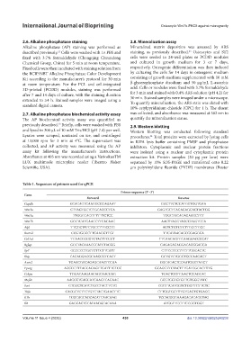
Table 1. Sequences of primers used for qPCR
Primer sequence (5’–3’)
Gene
Forward Reverse
Gapdh GCACAGTCAAGGCCGAGAAT GCCTTCTCCATGGTGGTGAA
Wnt3a CTTAGTGCTCTGCAGCCTGA GAGTGCTCAGAGAGGAGTACTGG
Wnt5a TTGGCCACGTTTTTCTCC TGGCTGCAGAGAGGCTGT
Wnt7b GCCTCATGAACCTTCACAAC AACTTAGGTAGCGTGGTCCA
Alpl TTCGCTATCTGCCTTGCCTG AGTCTGTGTCTTGCCTGCC
Runx2 CCGTGGCCTTCAAGGTTGT TTCATAACAGCGGAGGCA
Col1a1 TCAACCCCGTCTACTTCCCT TTCAACAGTCCAAGAACCCCAT
Bglap GCCTACAAACGCATCTACGG GAGAGAGAGGACAGGGAGGA
Sp7 GCCCCCTGGTGTTCTTCATT CTTCCCCCTTCTTGGCACTC
Ibsp CAGAGGAGGCAAGCGTCACT GCTGTCTGGGTGCCAACACT
Axin2 TGAGCGGCAGAGCAAGTCCAA GGCAGACTCCAATGGGTAGCT
Pparg AGCCCTTTACCACAGTTGATTTCTCC GCAGGTTCTACTTTGATCGCACTTTG
Cebpa TGGACAAGAACAGCAACGAG TCACTGGTCAACTCCAGCAC
Mef2c AAGCCTCAGCATCAAGTCAGAAC GCGTGGTGTGTTGTGGGTATC
Sost GTGCCTCATCTGCCTACTTGTG CGGTTCATGGTCTGGTTGTTCTC
Tnfa CACGCTCTTCTGTCTACTGAACTTC CTTGGTGGTTTGTGAGTGTGAGG
Il1b TCGCAGCAGCACATCAACAAG TCCACGGGAAAGACACAGGTAG
Il8 GAGCACTCCATAAGGCACAAA ATGGTTCCTTCCGGTGGT
Volume 11 Issue 4 (2025) 430 doi: 10.36922/IJB025240238