
Page 442 - v11i4
P. 442
International Journal of Bioprinting Osteocytic Wnt7b-PKCδ against microgravity
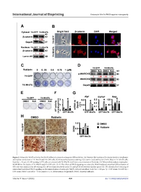
Figure 2. Osteocytic Wnt7b activates the PKCδ pathway to promote osteogenic differentiation. (A) Western blot analysis of β-catenin levels in cytoplasmic
and nuclear extractions of Y4-Wnt7b and Y4-GFP cells. (B) Immunofluorescence staining of β-catenin (red) and nuclei (DAPI, blue) in Y4-Wnt7b cells.
Scale bar = 50 µm. (C) AP staining in ST2 cells co-cultured with Y4-Wnt7b in different concentrations of Rottlerin. (D) Western blot analysis of phospho-
MARCKS in the lysates of Y4-Wnt7b and Y4-GFP cells. (E–G) The effect of PKCδ signaling on osteocytic Wnt7b-induced osteoblast differentiation of
ST2 cells was examined by AP staining (E), AP biochemical activity assay (F), and qPCR of osteoblast marker genes (G). (H) Alizarin Red S staining of
mineralized nodulesof PKCδ signaling on the mineralization of osteocytic Wnt7b-induced osteoblast. Scale bar = 100 µm. *p < 0.05 versus Y4-GFP, #p <
0.05 versus DMSO control (n = 3) for panels F, G, H. Abbreviations: Bright field; DMSO, dimethyl sulfoxide.
Volume 11 Issue 4 (2025) 434 doi: 10.36922/IJB025240238