
Page 443 - v11i4
P. 443
International Journal of Bioprinting Osteocytic Wnt7b-PKCδ against microgravity
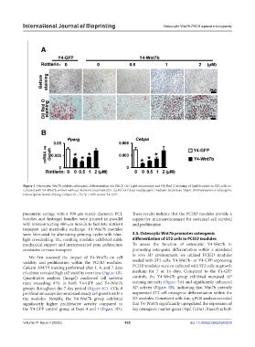
Figure 3. Osteocytic Wnt7b inhibits adipogenic differentiation via PKCδ. (A) Light microscopy and Oil Red O staining of lipid droplets in ST2 cells co-
cultured with Y4-Wnt7b, with or without Rottlerin treatment (0.5–2 µM) for 7 days in adipogenic medium. Scale bars: 50 µm. (B) Expression of adipogenic
transcription factors (Pparg, Cebpa) (n = 3); *p < 0.05 versus Y4-GFP.
pneumatic syringe with a 300-μm nozzle diameter. PCL These results indicate that the PCI3D modules provide a
bundles and hydrogel bundles were printed in parallel supportive microenvironment for sustained cell survival
with interconnecting 400-μm tunnels to facilitate nutrient and proliferation.
transport and metabolite exchange. Y4-Wnt7b modules
were fabricated by alternating printing cycles with blue- 3.5. Osteocytic Wnt7b promotes osteogenic
light crosslinking. The resulting modules exhibited stable differentiation of ST2 cells in PCI3D modules
mechanical support and interconnected pore architecture To assess the function of osteocytic Y4-Wnt7b in
conducive to mass transport. promoting osteogenic differentiation within a simulated
We first assessed the impact of Y4-Wnt7b on cell in vivo 3D environment, we utilized PCI3D modules
viability and proliferation within the PCI3D modules. seeded with ST2 cells. Y4-Wnt7b- or Y4-GFP-expressing
Calcein AM/PI staining performed after 1, 4, and 7 days PCI3D modules were co-cultured with ST2 cells in growth
of culture revealed high cell viability over time (Figure 4B). medium for 7 or 14 days. Compared to the Y4-GFP
Quantitative analysis (ImageJ) confirmed cell survival controls, the Y4-Wnt7b group exhibited increased AP
rates exceeding 97% in both Y4-GFP and Y4-Wnt7b staining intensity (Figure 5A) and significantly enhanced
groups throughout the 7-day period (Figure 4C). CCK-8 AP activity (Figure 5B), indicating that Wnt7b potently
proliferation assays demonstrated steady cell growth within augmented ST2 cell osteogenic differentiation within the
the modules. Notably, the Y4-Wnt7b group exhibited 3D modules. Consistent with this, qPCR analysis revealed
significantly higher proliferative activity compared to that Y4-Wnt7b significantly upregulated the expression of
the Y4-GFP control group at Days 4 and 7 (Figure 4D). key osteogenic marker genes (Alpl, Col1a1, Runx2) at both
Volume 11 Issue 4 (2025) 435 doi: 10.36922/IJB025240238