
Page 60 - v11i4
P. 60
International Journal of Bioprinting 3D bioprinting of nerve guidance conduits
direction of nerve cells, promoting cell proliferation conduits with excellent structural integrity, such as neural
during nerve tissue regeneration. Wu et al. used DLP to conduits, can be fabricated. Extrusion 3D printing offers
157
prepare an active hydrogel composed of GelMA and SF high deposition through (multi) nozzles at high printing
methacrylate, which had a positive effect on the adhesion, speeds and low equipment costs. However, the system is
proliferation, and migration of SCs. A 12 mm sciatic prone to nozzle clogging and pressure drops, which may
nerve defect model was established in rats, and a conduit eventually lead to cell apoptosis. 160,161
was implanted. Electrophysiological, morphological, and Vijayavenkataraman et al. developed an extrusion-
162
histological evaluations demonstrated that the conduit based 3D printing machine to fabricate NGCs using a
could promote axonal regeneration, myelin sheath conductive collagen/PPy-block-PCL hydrogel. The authors
regeneration, and functional restoration by providing a investigated the effect of printing speed and material flow
favorable microenvironment, with strong potential for the rate on the printed path width (Figure 8A), demonstrating
treatment of long-gap peripheral nerve injuries.
that, as the print speed increases, the material is subjected
4.3. Extrusion printing to tension, and the path width narrows or is discontinued.
Extrusion printing is the most widely used 3D printing At higher material flow rates, the ink may also aggregate
technique in NTE. The material is loaded and continuously into irregular shapes rather than a continuous line,
extruded from the print head through a nozzle and affecting the structure of the NGC. Although low-viscosity
selectively deposited LBL according to a pre-generated materials are often a good choice for maintaining cell
path, 158,159 with an acceptable resolution between 200 viability during and after printing, high-viscosity materials
and 2000 μm. Extrusion printing allows the deposition can provide better support and fidelity during the printing
of biomaterials with relatively high viscosity and shear- process. Therefore, the right trade-off between printing
thinning effect, which withstands during the printing speed and flow rate was demonstrated to be critical. Kaplan
process, preventing any collapse. Therefore, porous et al. also printed poly(L-lactic acid)/PLGA conduits
163
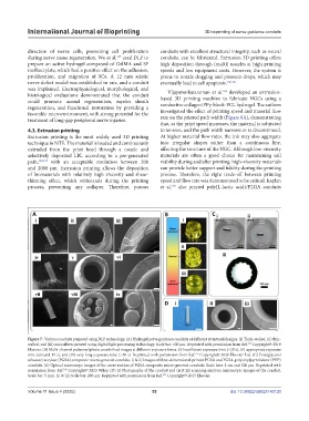
Figure 7. Various conduits prepared using DLP technology. (A) Hydrogel nerve guidance conduits of different structural designs. (i) Thick-walled, (ii) thin-
walled, and (iii) microfibers printed using digital light processing technology. Scale bar: 100 μm. Reprinted with permission from Ref. Copyright© 2019
153
Elsevier. (B) Multi-channel patterned photo crosslinked images at different exposure times. (i) Insufficient exposure time (<20 s), (ii) appropriate exposure
time (around 15 s), and (iii) very long exposure time (>50 s). Reprinted with permission from Ref. Copyright© 2020 Elsevier Ltd. (C) Poly(glycerol
155
sebacate) acrylate (PGSA) composite micro-grooved conduits. (i & ii) Images of three-dimensional-printed PGSA and PGSA-polyvinylpyrrolidone (PVP)
conduits. (ii) Optical microscope images of the cross-section of PGSA composite micro-grooved conduits. Scale bars: 1 cm and 200 μm. Reprinted with
permission from Ref. Copyright© 2023 Wiley. (D) (i) Photographs of the conduit and (ii & iii) scanning electron microscopic images of the conduit.
156
Scale bar: 5 mm. (ii & iii) Scale bar: 200 μm. Reprinted with permission from Ref. Copyright© 2015 Elsevier.
157
Volume 11 Issue 4 (2025) 52 doi: 10.36922/IJB025140120