
Page 63 - v11i4
P. 63
International Journal of Bioprinting 3D bioprinting of nerve guidance conduits
reported NGC prototypes have all been extensively variety of medical applications, including PLA, PLGA, and
tested in specific case studies, which validated their collagen. For instance, NeuraGen® and NeuroMatrix® are
efficient use. Nevertheless, their exploitation and clinical Food and Drug Administration-approved nerve conduits
translation remain limited. Several key challenges must made from collagen type I and primarily used for small-
be addressed before NGCs can be widely adopted in diameter peripheral nerves with short defects. Particularly,
preclinical and clinical studies. These challenges are mainly NeuraGen® is a tube filled with chondroitin-6-sulfate,
related to the selection of appropriate biomaterials and which promotes SC migration and axonal regeneration.
technologies, as highlighted in the previous sections. For Their functionality was demonstrated in rat sciatic nerve
biomaterials, European Medicines Agency or Food and gap models to increase nerve fiber density and myelinated
Drug Administration approval is a critical requirement. axon counts, and was comparable to reversed autografts
Several biomaterials have already been approved for a and superior to hollow or collagen-filled conduits.
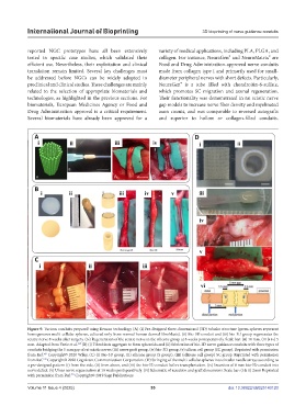
Figure 9. Various conduits prepared using Kenzan technology (A) (i) Pre-designed three-dimensional (3D) tubular structure (green spheres represent
homogeneous multi-cellular spheres, cultured only from normal human dermal fibroblasts). (ii) Bio-3D conduit and (iii) bio-3D group regenerates the
sciatic nerve 8 weeks after surgery. (iv) Regeneration of the sciatic nerve in the silicone group at 8 weeks postoperatively. Scale bar: (ii) 10 mm. (iii & iv) 5
168
mm. Adapted from Yurie et al. (B) (i) Fibroblasts aggregate to form spheroids and (ii) fabrication of bio-3D nerve guidance conduits with three types of
conduits bridging the 5 mm gap of rat sciatic nerve: (iii) nerve graft group, (iv) bio-3D group, (v) silicon cell group (SC group). Reprinted with permission
169
from Ref. Copyright© 2020 Wiley. (C) (i) Bio-3D group, (ii) silicone group (S group), (iii) (silicone cell group) SC group. Reprinted with permission
170
from Ref. Copyright© 2020 Cognizant Communication Corporation. (D) Stringing of the multi-cellular spheres into circular needle arrays according to
a pre-designed pattern (i) from the side, (ii) from above, and (iii) the bio-3D conduit before transplantation. (iv) Insertion of 8 mm bio-3D conduit into
nerve defect. (v) Ulnar nerve regeneration at 10 weeks postoperatively. (vi) Schematic of resection and graft dimensions. Scale bar: (i & ii) 2mm Reprinted
171
with permission from Ref. Copyright© 2019 Sage Publications.
Volume 11 Issue 4 (2025) 55 doi: 10.36922/IJB025140120