
Page 57 - IMO-2-3
P. 57
Innovative Medicines & Omics Biocompatibility of nanomaterials
biocompatibility. In cell culture studies using osteoblasts,
these scaffolds supported healthy cell growth, attachment,
and differentiation, with minimal toxicity. The controlled
release of calcium and phosphate ions also encouraged
robust matrix mineralization. 42
These findings were backed by in vivo experiments in
rodent models, where CaO–CaP scaffolds were implanted
into critical-size bone defects. Tissue analysis showed
strong new bone formation, seamless integration with
host tissue, and tight bonding at the interface. Compared
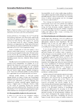
Figure 1. Diagram illustrating the material properties and molecular to conventional grafts, CaO–CaP composites accelerated
mechanisms of the CaO–CaP binary system. Image created by the author. healing and enhanced defect closure, reinforcing their
Abbreviations: CaO: Calcium oxide; CaP: Calcium phosphate.
suitability for clinical use. 43,44
38
elevate performance. For instance, Qi et al. found that 6.4. Clinical bottlenecks and inflammation response
incorporating magnesium into bioceramics significantly
improved cell response and vascular development, both Despite their clear advantages, CaO–CaP scaffolds face
essential for bone healing. This supports the growing some hurdles in clinical translation, particularly regarding
view that finely tuned ion release—including calcium, inflammation caused by rapid degradation. The high
phosphate, and magnesium ions—helps replicate the bone’s dissolution rate of CaO can cause spikes in calcium
natural healing environment and bolsters the rationale for ion levels and increase local pH, which may irritate
materials such as CaO–CaP in regenerative design. surrounding tissues and trigger immune responses.
Another valuable trait of CaO–CaP systems is their In our own pre-clinical tests, areas where the scaffold
inherent antimicrobial potential, increasingly important degraded quickly showed signs of local inflammation
for reducing post-surgical infection risks. The basic nature and mild immune cell activation—likely a response to
of CaO elevates the surrounding pH upon dissolution, sudden changes in ion concentration and pH. To address
disrupting bacterial membranes, denaturing proteins, and this, we applied biodegradable polymer coatings such
impairing enzyme function, ultimately killing harmful as poly(lactic-co-glycolic acid) (PLGA) and PEG to the
microbes. 39 scaffold surface. These coatings help regulate the ion release
profile, minimize pH shifts, and reduce inflammatory
This effect has been demonstrated using CaO and calcium responses. Our findings align with prior research showing
peroxide (CaO ) nanoparticles. For example, Yu et al. that surface modification of CaO-based materials can
40
2
reported that polyacrylic acid-coated CaO nanoparticles delay degradation and mitigate adverse reactions while
2
not only supported wound healing but also combated maintaining regenerative function. 45,46 As illustrated in
bacterial growth through the combined release of calcium Figure 2, PLGA-coated CaO–CaP scaffolds appear to
ions and reactive oxygen species. Similarly, Levingstone activate a more controlled immune response, particularly
et al. found that CaP-based scaffolds not only promoted in relation to cytokine release.
41
bone regeneration but also resisted bacterial colonization.
Further analysis of the inflammatory microenvironment
Although specific research on CaO–CaP systems revealed that early-stage responses (0–7 days post-
remains nascent, our team at Science and Technological implantation) were characterized by elevated levels of
Enhanced Laboratory for Advanced Learning and Research pro-inflammatory cytokines such as IL-6, TNF-α, and
(S.T.E.L.L.A.R) Laboratories is actively evaluating their IL-1 beta. These mediators contribute to tissue swelling,
antibacterial potential—especially against Staphylococcus leukocyte recruitment, and vascular changes. In the
aureus, a frequent cause of orthopedic infections. Initial subacute phase, the inflammatory signal begins to subside,
in vitro results are promising, showing less bacterial making way for reparative processes. A critical aspect of
adhesion and better scaffold sterility. This points to the dual recovery is the phenotypic transition of macrophages
functionality of CaO–CaP materials: supporting tissue repair from the pro-inflammatory M1 phenotype to the anti-
while simultaneously offering protection against infection. inflammatory M2 phenotype. This shift is associated with
increased secretion of anti-inflammatory cytokines like
6.3. Biocompatibility studies (in vitro and in vivo)
IL-10, which downregulate the immune response and
A series of in vitro and in vivo evaluations confirms promote tissue regeneration. Modulating this immune
that CaO–CaP nanomaterials exhibit excellent balance through material design and surface treatment is
Volume 2 Issue 3 (2025) 51 doi: 10.36922/IMO025210024