
Page 58 - IMO-2-3
P. 58
Innovative Medicines & Omics Biocompatibility of nanomaterials
key to minimizing long-term tissue damage and enhancing Beyond biochemical cues, gene-activated scaffolds have
scaffold integration. As shown in Figures 3 and 4, PLGA emerged as a powerful tool, enabling the localized delivery
modification alters the functional behavior of CaO–CaP of therapeutic DNA or RNA to stimulate regenerative
nanomaterials, while subsequent scaffold degradation pathways directly within the defect area. These platforms
triggers a time-dependent inflammatory response marked are gaining attention for their potential in treating complex
by shifts in cytokine expression. and non-healing bone injuries, where conventional
6.5. Emerging strategies for clinical translation scaffolds often fall short.
Translating CaO–CaP-based systems into clinical A transformative shift is also underway with the adoption
practice requires more than just demonstrating biological of 3D printing technologies, allowing the fabrication
compatibility; it also demands innovations in material of patient-specific scaffolds with precise anatomical
design, delivery mechanisms, and scalable fabrication conformity. This level of personalization improves not only
methods. One notable advancement is the inclusion of implant integration and mechanical performance but also
osteogenic growth factors such as bone morphogenetic healing outcomes. As part of our ongoing investigations,
proteins (BMPs) and vascular endothelial growth factor CaO–CaP scaffolds are being combined with bioactive
(VEGF), which play key roles in promoting both osteoblast molecules and additive manufacturing techniques to
differentiation and vascularization of the implant site. 47 boost regenerative efficiency while enhancing clinical
adaptability. 48
In parallel, ion-doped biodegradable systems—
particularly those incorporating magnesium ions—are
showing great promise for bone repair. A recent study
by Tao et al. described the successful development of
49
porous PLA-based microspheres doped with magnesium,
which exhibited enhanced biocompatibility, improved
osteogenic potential, and controlled biodegradation.
Figure 2. A series of reactions triggered by the surface modification of These findings align with our own data, underscoring
CaO–CaP. Adding PLGA coating changes the pH and affects the secretion the importance of controlled ionic release and scaffold
of pro-inflammatory cytokines. Image created by the author.
Abbreviations: CaO: Calcium oxide; CaP: Calcium phosphate; adaptability in the clinical success of CaO–CaP
PLGA: Poly(lactic-co-glycolic acid). materials.
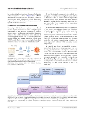
Figure 3. The effect of PLGA modification on the behavior of CaO–CaP nanomaterials. The uncoated pathway results in rapid calcium ion release and pH
elevation, which may lead to tissue irritation and upregulation of inflammatory cytokines such as IL-6 and TNF-α. In contrast, the PLGA-coated pathway
moderates ion release and stabilizes pH, thereby reducing inflammation and improving biocompatibility. Image created by the author.
Abbreviations: CaO: Calcium oxide; CaP: Calcium phosphate; IL-6: Interleukin-6; PLGA: Poly(lactic-co-glycolic acid); TNF-α: Tumor necrosis
factor alpha.
Volume 2 Issue 3 (2025) 52 doi: 10.36922/IMO025210024