
Page 30 - ITPS-6-2
P. 30
INNOSC Theranostics and
Pharmacological Sciences Residual versus curative antimalarial tests
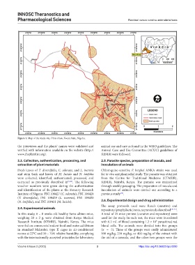
Figure 1. Map of the study site, Omu-Aran, Kwara State, Nigeria.
the interviews and the plants’ names were validated and animal use and care as found in the WHO guidelines. The
verified with information available on the website (http:// Animal Care and Use Committee (ACUC) guidelines of
www.theplantlist.org). KEMRI were followed.
2.3. Collection, authentication, processing, and 2.5. Parasite species, preparation of inocula, and
extraction of plant materials inoculation of animals
Fresh leaves of T. diversifolia, C. odorata, and L. inermis Chloroquine-sensitive P. berghei ANKA strain was used
and stem bark and leaves of M. lucida and N. latifolia for in vivo antiplasmodial study. The parasite was obtained
were collected, identified, authenticated, processed, and from the Centre for Traditional Medicine (CTMDR),
extracted as previously described in [9,10] . The following KEMRI, Nairobi, Kenya. The parasite was maintained
voucher numbers were given during the authentication through weekly passaging. The preparation of inocula and
and identification of the plants at the Forestry Research inoculation of animals were carried out according to a
Institute of Nigeria: FNI 108427 (C. odorata), FNI 108428 previous study .
[13]
(T. diversifolia), FNI 108429 (L. inermis), FNI 108430
(N. latifolia), and FNI 108431 (M. lucida). 2.6. Experimental design and drug administration
The assay protocols used were Rane’s (curative) and
2.4. Experimental animals repository (prophylactic) tests, as previously described [20-22] .
In this study, 6 – 8 weeks old healthy Swiss albino mice, A total of 25 mice per test (curative and repository) were
weighing 20 ± 2 g, were obtained from Kenya Medical used for the study. In each test, the mice were inoculated
Research Institute (KEMRI), Nairobi, Kenya. The mice with 0.2 mL of blood containing 1.2 × 10 parasitized red
7
were fed on commercial rodent food and water ad libitum blood cells. The animals were divided into five groups
in standard Makrolon type II cages in air-conditioned (n = 5). Three of the groups were orally administered
rooms at 22°C and 50 – 70% relative humidity, complying 100 mg/kg, 250 mg/kg, or 400 mg/kg of the extract with
with the internationally accepted principles for laboratory the aid of a cannula, and the other two groups were the
Volume 6 Issue 2 (2023) 3 https://doi.org/10.36922/itps.0300