
Page 47 - ITPS-6-2
P. 47
INNOSC Theranostics and
Pharmacological Sciences Genotoxicity of (4-fluorophenyl) thiazolidin-4-one
safety assessment is essential for their further clinical
development . Therefore, in this study, we conducted a
[34]
detailed cytogenetic investigation on 4-TH using a normal
mammalian cell line.
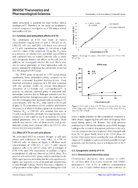
3.1. Cytotoxic and antioxidant effects of 4-TH
The cytotoxicity of 4-TH was tested at various
concentrations ranging from 0.1 µM to 100 µM on the
CHO-K1 cell line, and 50% cell death was observed
at 7.5 µM concentration (Figure 1), indicating a high
cytotoxic nature of the compound. Using this IC value
50
as a reference, we selected three sub-lethal concentrations
and conducted tests on normal CHO-K1 cells to assess Figure 1. Percentage of cytotoxic effect (MTT assay) of 4-TH on CHO-
their cytogenetic toxicity and effects on the cell cycle. In K1 cell line.
addition, we investigated whether the toxic effects were
due to radical generation or direct interaction with the
genetic material by evaluating the antioxidant properties
of 4-TH using the DPPH assay.
The DPPH assay conducted on 4-TH demonstrated
significantly lower antioxidant activity compared to the
standard compound butylated hydroxytoluene. These
observed antioxidant properties of 4-TH were consistent
with the previous results on various thiazolidinone
derivatives of 1,3-thiazole and 1,3,4-thiadiazole . In
[35]
general, the phenolic hydroxyl group is associated with
antioxidant activities due to hydrogen donation and free
radical stabilization through resonance. The results showed
that the scavenging effect increased with increasing 4-TH
concentrations, with the EC value found to be 50 µM
50
(Figure 2). The antioxidant activity could be attributed to Figure 2. Antioxidant activity of 4-TH. Data are presented as the mean
the presence of phenolic hydroxy groups in the structures. of two independent replications ± standard error of the mean (SE); EC
50
However, IC in CHO-K1 cells revealed that even at value of 4-TH = 50 µM.
50
7.5 µM, it induced 50% cell death. This high toxicity in the
presence of 4-TH could be due to an increase in oxidant phase, a higher number of cells accumulated compared to
radical generation, even at low concentrations. These other phases, suggesting that cells were undergoing DNA
radicals may interact with cell biomolecules and genetic repair during mitotic cell division. This study provides
material, causing more cytotoxic effects in CHO-K1 cells, evidence that 4-TH has the ability to induce DNA damage,
even at very low concentrations. leading to the activation of apoptosis pathways in the cells.
It is also assumed that the unrepaired DNA-damaged cells
3.2. Effect of 4-TH on cell cycle phases
from the G1 phase finally arrest at the G2/M phase for
We performed FACS to evaluate changes in cell cycle further DNA repair before entering the next cell division.
phases and understand the toxic effect of 4-TH on Hence, 4-TH is likely to induce cell death, possibly through
CHO-K1 cells. As shown in Figure 3, all three tested apoptosis, and the toxicity caused is independent of the S
concentrations of 4-TH (2.5, 5, and 7.5 µM) induced phase.
apoptotic cells in the sub-G1 phase, with percentages of
8.7%, 5.9%, and 7.5%, respectively. In addition, a higher 3.3. Cytogenetic toxicity of 4-TH
accumulation of cells was observed in G0/G1 phase. This 3.3.1. Chromosomal aberration assay
arrest of cells at the G0/G1 phase could be attributed to the
DNA repair process, indicating that the cells experienced a Chromosomal aberrations were assessed in CHO-
highly toxic exposure to the compound and required time K1 cell lines after 24 h of post-treatment with DMSO,
to pass through S-phase. Thereby there was a reduction in mitomycin-C, and 4-TH at different concentrations
cell population in the S phase. Moreover, during the G2/M (2.5, 5, and 7.5 µM). DMSO-treated cells exhibited 26
Volume 6 Issue 2 (2023) 4 https://doi.org/10.36922/itps.0618