
Page 50 - ITPS-6-2
P. 50
INNOSC Theranostics and
Pharmacological Sciences Genotoxicity of (4-fluorophenyl) thiazolidin-4-one
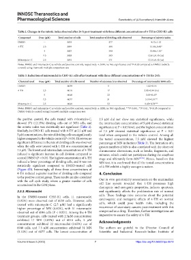
Table 2. Changes in the mitotic index observed after 24‑h post‑treatment with three different concentrations of 4‑TH in CHO‑K1 cells
Compound Dose (µM) Total number of cells Total number of dividing cells observed Percentage of mitotic index
DMSO -- 3035 277 9.12±1.12
4-TH 2.5 3007 406 13.50±0.60*
5 3027 390 12.88±1.33*
7.5 3028 383 12.64±0.45 (ns)
Mitomycin- C 2.5 3051 371 12.15±0.42 (ns)
Notes: DMSO and mitomycin-C as vehicle and positive controls, respectively. ±: SEM; ns: Not significance and *P<0.05 compared to DMSO (vehicle
control) using Dunnett’s multiple comparison test
Table 3. Induction of micronuclei in CHO‑K1 cells after treatment with three different concentrations of 4‑TH for 24 h
Chemical used Dose (µM) Total number of cells scored Number of micronucleus observed Percentage of micronuclei/1000 cells
DMSO - 6039 11 1.82±0.16
4-TH 2.5 6016 17 2.82±0.34 (ns)
5 6048 22 3.63±0.43*
7.5 6077 31 5.10±0.31**
Mitomycin-C 2.5 6046 52 8.60±0.70***
Notes: DMSO and mitomycin-C as vehicle and positive controls, respectively. ±: SEM; ns: Not significant, ***P<0.001, **P<0.01, *P<0.05 compared to
DMSO (vehicle control) using Dunnett’s multiple comparison test.
the positive control, the cells treated with mitomycin-C, 2.5 µM did not show any statistical significance, while
showed 371 (12.15%) dividing cells out of 3051 cells, and the intermediate concentration of 5 µM showed statistical
the mitotic index was statistically not significant (Table 2). significance at P < 0.05 level, and the highest concentration
Similarly, in CHO-K1 cells treated with 4-TH at 2.5 µM and of 7.5 µM showed statistical significance at P < 0.01
5 µM concentrations, the rate of dividing cells was significantly level when compared to the vehicle control. Among all
higher compared to the vehicle control (P < 0.05). However, no the tested concentrations, 7.5 µM showed the highest
significant difference in the rate of dividing cells was observed percentage of MN induction (Table 3). The formation of a
when the cells were treated with 4-TH at a concentration of greater number of MN is also correlated with the observed
7.5 µM. The lowest and intermediate concentrations of 4-TH chromosome aberrations, such as breaks, fragments, and
induced a significant increase in cell division compared to minutes, which could not participate during the anaphase
control DMSO (P < 0.05). The highest concentration of 4-TH stage and ultimately form MN [37,38] . Hence, based on this
induced a lower percentage of dividing cells, and it was not MN test, it is confirmed that all the tested concentrations
statistically significant compared to DMSO-treated cells of 4-TH exhibit a highly aneugenic nature.
(Figure 4D). Interestingly, all these three concentrations of
4-TH induced a greater number of dividing cells compared 4. Conclusion
to the positive control group. These results are also correlated Our in vitro genotoxicity assessments on the mammalian
with the cell cycle study, where a greater number of cells
accumulated in the G/M phase. cell line system revealed that 4-TH possesses high
clastogenic and aneugenic properties, induces apoptosis,
3.3.3. Micronuclei and significantly affects the proliferation rate of normal
In the DMSO-treated CHO-K1 cells, 11 micronuclei cells. These findings raise concerns about the potential
(1.82%) were observed out of 6039 cells. However, cells carcinogenic and mutagenic effects of 4-TH on normal
treated with mitomycin-C (2.5 µM) had a significantly cells, which could pose health risks, including the
higher percentage of MN (8.60%), with 51 micronuclei recurrence of secondary cancers post-treatment with this
observed out of 6046 cells (P < 0.001). Among the 4-TH compound as a drug. Therefore, further investigations are
treatment groups, cells treated with 2.5µM concentration imperative to ensure the safety of 4-TH.
exhibited 17 MN (2.82%) out of 6016 cells, 5 µM Acknowledgments
concentration exhibited 22 micronuclei (3.63%) out of
6048 cells, and 7.5 µM concentration exhibited 31 MN The authors are grateful to the Director Council of
(5.10%) out of 6077 cells. The lowest concentration of Scientific and Industrial Research-Indian Institute of
Volume 6 Issue 2 (2023) 7 https://doi.org/10.36922/itps.0618