
Page 40 - ITPS-8-2
P. 40
INNOSC Theranostics and
Pharmacological Sciences MDD biomarkers: Clinical implications
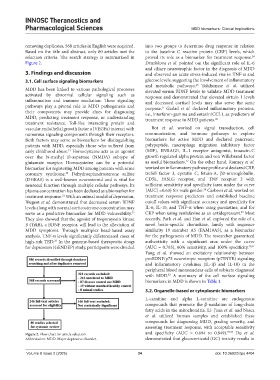
removing duplicates, 568 articles in English were acquired. into two groups to determine drug response in relation
Based on the title and abstract, only 80 articles met the to the baseline C reactive protein (CRP) levels, which
selection criteria. The search strategy is summarized in proved its role as a biomarker for treatment response.
24
Figure 2. Druzhkova et al. pointed out the significant role of IL-6
and ciliary neurotrophic factor in the diagnosis of MDD
3. Findings and discussion and observed an acute stress-induced rise in TNF-α and
3.1. Cell surface signaling biomarkers glucose levels, suggesting the involvement of inflammatory
and metabolic pathways. Tolahunase et al. utilized
25
MDD has been linked to various pathological processes elevated serum BDNF levels to validate MDD treatment
activated by abnormal cellular signaling such as response and demonstrated that elevated sirtuin 1 levels
inflammation and immune mediation. These signaling and decreased cortisol levels may also serve the same
pathways play a pivotal role in MDD pathogenesis and purpose. Gadad et al. declared inflammatory proteins,
21
their components may provide clues for diagnosing i.e., interferon-gamma and eotaxin/CCL1, as predictors of
MDD, predicting treatment response, or understanding treatment response in MDD patients. 26
treatment resistance. Toll-like interacting protein and
vascular endothelial growth factor a (VEGFa) interact with Bot et al. worked on signal transduction, cell
numerous signaling components through their receptors. communication, and immune pathways to explore
Both factors may serve as biomarkers for distinguishing biomarkers for active MDD and declared pancreatic
patients with MDD, especially those who suffered from polypeptide, macrophage migration inhibitory factor
early childhood abuse. Homocysteine acts as an agonist (MIF), ENRAGE, IL-1 receptor antagonist, tenascin-C
19
over the N-methyl D-aspartate (NMDA) subtype of growth regulated alpha protein and von Willebrand factor
27
glutamate receptor. Homocysteine can be a potential as useful biomarkers. On the other hand, Ramsey et al.
biomarker for registering MDD among patients with acute worked on inflammatory pathway profile and declared CRP,
coronary syndrome. Dehydroepiandrosterone sulfate trefoil factor 3, cystatin C, fetuin-A, β2-microglobulin,
20
(DHEAS) is a well-known neurosteroid and is vital for CD5L, FASLG receptor, and TNF receptor 2 with
neuronal function through multiple cellular pathways. Its sufficient sensitivity and specificity (area under the curve
28
plasma concentration has been declared as a biomarker for [AUC] =0.63) for male gender. Carboni et al. worked on
treatment response. With an animal model of depression, treatment response predictors and established baseline
21
Blugeot et al. demonstrated that decreased serum BDNF cutoff values with significant accuracy and specificity for
levels along with normal corticosterone concentration may IL-6, IL-10, and TNF-α when using paroxetine, and for
29
serve as a predictive biomarker for MDD vulnerability. CRP when using venlafaxine as an antidepressant. Most
22
They also showed that the agonist of tropomyosin kinase recently, Park et al. and Han et al. explored the role of a
B (TrkB), a BDNF receptor, will lead to the alleviation of novel brain-specific chemokine, family with sequence
MDD symptoms. Through multiplex bead-based assay similarity 19 member A5 (FAM19A5), as a biomarker
analysis, TNF-α levels significantly differentiated cases of for the pathogenesis of MDD. The researcher guaranteed
high-risk TRD. In the genome-based therapeutic drugs authenticity with a significant area under the curve
23
for depression (GENDEP) study, participants were divided (AUC = 0.785), 60% sensitivity, and 100% specificity. 30,31
Yang et al. showed an excitatory relationship between
proBDNF/p75 neurotropic receptors (p75NTR) signaling
and inflammatory cytokines (IL-1β and IL-10) in the
peripheral blood mononuclear cells of subjects diagnosed
with MDD. A summary of the cell surface signaling
32
biomarkers in MDD is shown in Table 1.
3.2. Organelle-based or cytoplasmic biomarkers
L-carnitine and alpha L-carnitine are endogenous
compounds that promote the β-oxidation of long-chain
fatty acids in the mitochondria. Li- Juan et al. and Nasca
et al. utilized human samples and established these
compounds for diagnosing MDD, grading severity, and
assessing treatment response, with acceptable sensitivity
Figure 2. Flow chart for article selection and specificity (AUC = 0.694 to 0.849). 33,34 Du et al.
Abbreviation: MDD: Major depressive disorder. demonstrated that glucocorticoid (GC) toxicity results in
Volume 8 Issue 2 (2025) 34 doi: 10.36922/itps.4404