
Page 22 - JCTR-10-5
P. 22
284 Cirrincione | Journal of Clinical and Translational Research 2024; 10(5): 283-290
and symmetrically pleasing boosts confidence in social
interactions and enhances attractiveness [1]. Dentistry has
adapted and improved its techniques to meet these new
requirements, strengthening the relationship between various
dental disciplines, such as prosthetics, orthodontics, and
implantology. This interdisciplinary connection has been
facilitated by the impressive development of digital techniques
in recent years. This includes the ability to easily manipulate
patient impressions obtained with intraoral scanners, such as
standard tessellation language (STL) files, and to combine them
with 3D visualization of the bone from digital imaging and
communications in medicine (DICOM) files generated by cone-
beam computed tomography (CBCT) [2]. Therefore, dentists
now have the opportunity to create a “virtual patient” on their
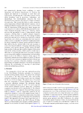
computers, allowing them to establish adequate diagnostic Figure 1. Maryland-type temporary composite bridge in site 12
criteria to obtain excellent results [3]. This strategy becomes
particularly important when operators are faced with a reduced
buccal bone wall, which can compromise the final long-term
esthetic results in immediate [4] and late [5] implant placement.
In contrast, adopting this strategy requires operators to improve
their skills to become familiar with all the tools necessary to
achieve the desired results [6]. Designing esthetically pleasing
prosthetic work requires absolute synergy among all dental
team members. In the past, this workflow required collaboration
between the various operators, which could be complex due to
difficulties in visualizing the final result. Conversely, the digital
process has greatly simplified communication between dentists, Figure 2. Palatal view detail of the bridge attached to teeth 11 and 13
thanks in part to the ability to visualize various steps in 3D,
especially in clinical cases where esthetics is critical [7]. The aim
of this work was to present an implant-prosthetic clinical case
resulting from a previous orthodontic treatment, successfully
treated using new digital technologies. This article was prepared
following the strengthening the reporting of observational
studies in epidemiology guidelines.
2. Methods
This retrospective clinical case was conducted according
to the 1964 Helsinki Declaration principles for biomedical
research involving human subjects. The patient was informed
of the nature of the study, its benefits, risks, and possible
alternative treatments, and written consent was also obtained
for the use of clinical images. The patient was a 22-year-old
man who complained of esthetic problems that arose after
a previous orthodontic treatment. The orthodontic therapy
involved reopening the space for tooth 12 to resolve agenesis Figure 3. Frontal view of the smile before treatment
of the related permanent element, along with a temporary
composite reconstruction of the conoid tooth 22. The intraoral but the root axes of teeth 13 and 11 converged toward the apices,
examination displayed a composite Maryland bridge replacing making traditional implant surgery difficult (Figures 8-10). The
tooth 22 (Figures 1 and 2), temporarily positioned by the anteroposterior view of the conoid dental element 22 displayed
orthodontist, probably in view of the implant therapy. Teeth 11 composite reconstruction with a large horizontal over-contour,
and 21 featured some old composite reconstructions, discordant most likely to compensate for the vestibulo-palatal inclination
coronal axes, and the presence of a diastema at the incisal level; of the root axis (Figure 11). The gingival parabolas of the upper
tooth 21 also appeared to be about 1 mm longer than tooth 11 anterior group appeared unlevelled. Furthermore, the patient
(Figures 3 and 4). In tooth 12, the CBCT (Promax 3D Max; had moderate gingival exposure. Hence, the proposed treatment
Planmeca, Finland) displayed an adequate vestibulo-palatal bone plan included the insertion of a small diameter implant in site
diameter for the insertion of a small-sized implant (Figures 5-7), 12 through computer-guided implant surgery, a zirconia crown
DOI: https://doi.org/10.36922/jctr.24.00035