
Page 24 - JCTR-10-5
P. 24
286 Cirrincione | Journal of Clinical and Translational Research 2024; 10(5): 283-290
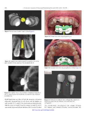
Figure 9. Apical view of teeth 13 and 11 (from Figure 8)
Figure 12. Frontal view of the surgical guide try-in
Figure 10. Sagittal cone-beam computed tomography view of the
space available for the insertion of the implant in site 12
Figure 13. Occlusal view of the surgical guide try-in (from Figure 12)
Figure 11. A sagittal view of the cone-beam computed tomography at
site 22, displaying the wide horizontal overcontour of the composite
reconstruction
digital impression was taken of both the temporary restoration Figure 14. Tooth 22 was extracted from coDiagnostix, imported to
adequately integrated into the soft tissues and the implant, as Meshmixer (figure above), mirrored, and re-imported into
well as teeth 11, 21, and 22. The final work was then delivered. coDiagnostix
Both the crown on tooth 22 and the veneers on teeth 11 and 21 AG, Liechtenstein), micro-layered with ceramic (Creation
were made of pressed lithium disilicate (MT; Ivoclar-Vivadent LS, Austria) and colored (Ivocolor; Ivoclar-Vivadent AG,
DOI: https://doi.org/10.36922/jctr.24.00035