
Page 25 - JCTR-10-5
P. 25
Cirrincione | Journal of Clinical and Translational Research 2024; 10(5): 283-290 287
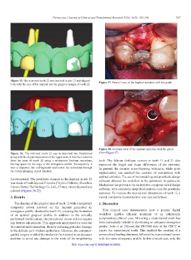
Figure 15. The mirrored tooth 22 was inserted in site 12 and aligned
both with the axis of the implant and the gingival margin of tooth 22 Figure 17. Frontal view of the implant insertion with the guide
Figure 18. Occlusal view of the implant insertion with the guide
Figure 16. The mirrored tooth 22 was re-imported into Meshmixer (from Figure 17)
along with the digital impression of the upper arch; it was then removed
from the zone of tooth 12 using a subtractive Boolean procedure, teeth. The lithium disilicate veneers in teeth 11 and 21 also
leaving space for the copy of the emergence profile. Subsequently, it improved the length and shape differences of the elements.
was re-imported into coDiagnostix and sent to the technician through In general, the ceramic micro-layering technique, while quite
the virtual planning export function.
sophisticated, has enabled the creation of restorations with
optimal esthetics. The use of two modeling and prosthetic design
Liechtenstein). The prosthetic element on the implant in site 12 software allowed the workflow to be optimized. In particular,
was made of multilayered Zirconia (Explore Esthetic; Shenzhen Meshmixer has proven to be an intuitive computer-aided design
Upcera Dental Technology Co. Ltd., China), micro-layered and software, with extremely simplified controls even for prosthetic
colored (Figures 20-22).
purposes. To increase the mesiodistal dimensions of tooth 13, a
3. Results mesial composite reconstruction was also performed.
The shaping of the gingival area of tooth 12 with a temporary 4. Discussion
composite crown screwed on the implant generated an
emergence profile identical to tooth 22, ensuring the formation This clinical case demonstrates how a precise digital
of an optimal gingival profile. In addition to the virtually workflow enables efficient treatment of an esthetically
performed modifications, the provisional crown did not require compromised clinical case. Mirroring a contralateral tooth has
any further adjustments. This approach minimized the need for been particularly helpful in developing an adequate emergence
its removal and reinsertion, thereby reducing potential damage profile. Joda et al. [8] used the DICOM data of the CBCT to
to the delicate peri-implant epithelium. Likewise, the computer- mirror the contralateral tooth. This enabled the creation of a
guided surgery enabled the implant to be inserted in an adequate personalized healing abutment, followed by a provisional crown
position to avoid any damage to the roots of the neighboring with the same emergency profile. In this clinical case, only the
DOI: https://doi.org/10.36922/jctr.24.00035