
Page 80 - JCTR-11-1
P. 80
Journal of Clinical and
Translational Research Treatment choice for iatrogenic A-dissection
2. Case presentation Given the mechanism of injury and the likely absence
of an intra-aortic intimal tear (evidenced by contrast
A 79-year-old patient, who had undergone exclusion of a stagnation in the false lumen), conservative treatment
degenerative abdominal aortic aneurysm 5 years earlier using was initially chosen. Antihypertensive treatment with
a bifurcated bi-iliac endovascular prosthesis, was admitted metoprolol (75 mg/day) and amlodipine (10 mg/day) was
for elective complementary endovascular treatment of initiated.
a progressively developing aneurysm in the distal right
iliac artery (Figure 1). After the successful retrograde A follow-up CT scan 48 h later showed near-complete
advancement of a catheter through the right femoral artery, regression of the false lumen. The patient was extubated
several attempts to insert a percutaneous guiding catheter and transferred to rehabilitation 14 days after admission,
antegrade through the left axillary artery failed to reach the with an unremarkable clinical course (Figure 3A).
descending thoracic aorta. Subsequent contrast injection A subsequent CT scan after 1 month confirmed the near-
revealed stagnation of the contrast medium at the aortic arch complete regression of the false lumen (Figure 3B).
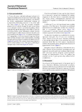
and ascending aorta (Figure 2A). A computed tomography Ten days later, the patient was readmitted due to acute,
(CT) angiography of the aorta showed a large false lumen oppressive chest pain. The CT scan now revealed the
filled with the previously used contrast medium, extending classic features of an acute aortic dissection, including
throughout the ascending aorta and into the distal aortic arch a free-moving intimal flap in the ascending aorta with
(Figures 2B and 2D). The contrast-delayed phase showed a persistent blood flow in both lumens (simultaneous
lack of contrast enhancement in this region, confirming an contrast enhancement) and an enlarged aortic diameter of
iatrogenic type A dissection (Figure 2C and 2E).
approximately 50 mm (Figure 4).
Emergency surgical replacement of the dissected aorta
with a Dacron vascular prosthesis (ascending aorta and
hemiarch) was performed under moderate hypothermia,
with continuous selective antegrade cerebral perfusion
through the brachiocephalic trunk. The patient was
extubated on the 1 post-operative day without further
st
complications.
3. Discussion
According to the European registry of iatrogenic type A
aortic dissections following cardiovascular procedures,
peripheral endovascular procedures account for only 1% of
all dissections reported. The registry comprises 18 centers
Figure 1. Aneurysm of the distal right iliac artery across eight European countries and includes 3902 patients
A B C
D E
Figure 2. Computed tomography angiography of the aorta. (A) Stagnation of contrast medium in the ascending aorta and the aortic arch after direct
injection in the left axillary artery catheter. (B and D) Native computed tomography scan shows a large false lumen extending throughout the ascending
aorta (B) and into the distal aortic arch (D), filled with the previously the previously injected contrast medium. (C and E) After intravenous contrast
medium injection, confirmation of an iatrogenic type A dissection.
Volume 11 Issue 1 (2025) 74 doi: 10.36922/jctr.24.00048