
Page 81 - JCTR-11-1
P. 81
Journal of Clinical and
Translational Research Treatment choice for iatrogenic A-dissection
A B mechanisms of injury (such as direct intimal damage to
the aorta caused by retrograde transaortic advancement of
guiding catheters or endoprostheses) differ from the case
presented here. In our case, the patient experienced a two-
6,7
stage clinical evolution. First, only a contrast-enhancing
aortic hematoma (without intimal tear) was observed,
C D for which conservative treatment was administered
without stenting of the entry point. This treatment was
initially successful, with complete regression of the
periaortic hematoma at 1 month. However, in the second
phase, an intimal tear developed despite well-managed
anti-hypertensive treatment, necessitating emergency
surgery for ascending aorta replacement. To the best of
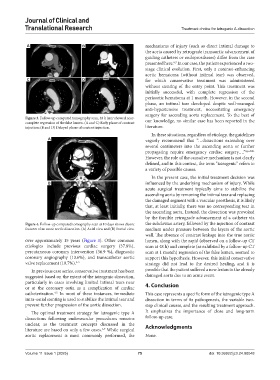
Figure 3. Follow-up computed tomography scan, 48 h later showed near- our knowledge, no similar case has been reported in the
complete regression of the false lumen. (A and C) Early phase of contrast
injection; (B and D) Delayed phase of contrast injection. literature.
In these situations, regardless of etiology, the guidelines
A vaguely recommend that “…dissections extending over
several centimeters into the ascending aorta or further
propagating require emergency cardiac surgery….” 8,p.2901
However, the role of the causative mechanism is not clearly
defined, and in this context, the term “iatrogenic” refers to
a variety of possible causes.
In the present case, the initial treatment decision was
B influenced by the underlying mechanism of injury. While
acute surgical treatment typically aims to stabilize the
ascending aorta by removing the intimal tear and replacing
the damaged segment with a vascular prosthesis, it is likely
that, at least initially, there was no corresponding tear in
the ascending aorta. Instead, the dissection was provoked
by the forcible retrograde advancement of a catheter via
Figure 4. Follow-up computed tomography scan at 10 days shows classic the subclavian artery, followed by the injection of contrast
features of an acute aortic dissection. (A) Axial view and (B) frontal view. medium under pressure between the layers of the aortic
wall. The absence of contrast leakage into the true aortic
over approximately 15 years (Figure 3). Other common lumen, along with the rapid (observed on a follow-up CT
etiologies include previous cardiac surgery (37.8%), scan at 48 h) and complete (as exhibited by a follow-up CT
percutaneous coronary intervention (36.9 %), diagnostic scan at 1 month) regression of the false lumen, seemed to
coronary angiography (13.6%), and transcatheter aortic support this hypothesis. However, this initial conservative
valve replacement (10.7%). 1-3 strategy did not lead to the desired healing, and it is
In previous case series, conservative treatment has been possible that the patient suffered a new lesion in the already
suggested based on the extent of the iatrogenic dissection, damaged aorta due to an acute event.
particularly in cases involving limited intimal tears near
or at the coronary ostia as a complication of cardiac 4. Conclusion
catheterization. In most of these instances, immediate This case represents a specific form of the iatrogenic type A
4,5
intra-ostial stenting is used to stabilize the intimal tear and dissection in terms of its pathogenesis, the variable two-
prevent further progression of the aortic dissection. step clinical course, and the resulting treatment approach.
The optimal treatment strategy for iatrogenic type A It emphasizes the importance of close and long-term
dissections following endovascular procedures remains follow-up care.
unclear, as the treatment concepts discussed in the Acknowledgments
literature are based on only a few cases. While surgical
6,7
aortic replacement is most commonly performed, the None.
Volume 11 Issue 1 (2025) 75 doi: 10.36922/jctr.24.00048