
Page 95 - JCTR-11-2
P. 95
Journal of Clinical and
Translational Research Biomimetic matrix used to treat diabetic foot ulcers
Table 1. Patients’ characteristics at the enrollment, including prior treatments, duration, wound location, wound depth, and
comorbidities prior to BMM treatment
S. no. Age (years) Prior treatments Wound duration (months) Wound location Wagner classification 14 Comorbidities
Patient 1 37 A, C, E, HA, LC, O, S, SX, V 39 Heel 2 D, N, OS
Patient 2 37 A, C, HA, LC, O, S, VS 30 Forefoot 1 N, OS, PVD
Patient 3 67 E, HA, O 8 Heel 2 D, N
Patient 4 58 A, C, E, HA, LC, LCA, O, S, SX, V 56 Forefoot 1 A, D, N, O, OS
Patient 5 88 C, E, HA, LC, LCA, S, V 3 Ankle 3 D, N, OS, PVD
Patient 6 69 O, S 15 Lateral Foot 1 C, D, N, PVD
Patient 7 70 A, C, E, HA, LC, LCA, O, S 8 Heel 3 D, N, O
Patient 8 84 C, HA, LCA, O, S 13 Mid-arch 2 C, N
Notes: Prior treatments: A: Amniotic membrane; C: Collagen-decellularized; E: Enzymatic debridement; H: Hyperbaric oxygen; HA: Hypochlorous
acid irrigation; LC: Living cell; LCA: Living cell amniotic; O: Off-loading shoe or boot; S: Silver dressing; SX: Surgery; V: Negative pressure wound
therapy; VS: Vascular surgery. Comorbidities: A: Partial amputation; C: Charcot deformity; D: Diabetes; N: Neuropathy; O: Obesity; OS: Osteomyelitis;
PVD: Peripheral vascular disease.
Abbreviation: BMM: Biomimetic matrix.
area. All measurements were taken following wound
debridement.
3. Results
This study selected a small clinical sample of eight patients
with stalled chronic diabetic foot wounds. The ulceration
duration before BMM treatment ranged from 3 up to
56 months (average = 21.5 months). The wounds were
located on weight-bearing surfaces of the foot, except for
one venous stasis ulcer located on the ankle in a patient who
was also diabetic. The wounds in this clinical case series
consisted of Wagner Grade 3 (n = 2; 25%), Grade 2 (n = 3;
37.5%), and Grade 1 (n = 3; 37.5%) ulcers. All wounds
had previously received standard of care, and, except for
one patient, wounds had also been previously treated with
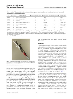
Figure 1. BMM is a transparent gel, and is provided in a sterile syringe. It multiple biologic products and failed to respond. Several
can be applied to the surface of the wound, or by inserting the flexible tip
into tunneling or undermined areas. wounds had also previously been treated with negative
Abbreviation: BMM: Biomimetic matrix. pressure therapy before treatment with BMM without
achieving significant improvement. A summary including
in place for 3 – 5 days, depending on the extent of drainage prior treatments, wound duration, wound location, wound
from the wound. The BMM was re-applied as needed for up depth, and comorbidities before BMM treatment of all the
to three applications per patient, and each application was subjects enrolled in this study can be found in Table 1.
normally 2 weeks apart. BMM is promptly integrated into All patients treated with BMM experienced a substantial
the tissue and bioresorbed, and therefore, does not need to reduction in the percent wound area, with an average
be removed from the wound site at dressing changes. decrease of 63.6%, ranging from 22% to 100% wound
All patients received appropriate off-loading to protect closure. Patients received one to three applications of
the wound site. Wounds were measured and photographed BMM. When considering similar wounds, it appears that
just before the initial application and at subsequent the number of applications of BMM does have an impact
visits. The wound area was calculated by tracing the on the percent change in wound area. For example, patients
wound perimeter to calculate the surface area. This was 1, 3, and 7 had similar comorbidities, and comparable
then compared to a standardized marker within the location (heel). However, patient 1 received only two
picture frame to calculate the wound surface area. These BMM treatments, while patients 3 and 7 each received
measurements were used to calculate changes in wound three treatments. Our data shows that patients 3 and 7 had
Volume 11 Issue 2 (2025) 89 doi: 10.36922/jctr.24.00063