
Page 9 - JCTR-11-3
P. 9
Journal of Clinical and
Translational Research Lateral patellar instability in deep flexion
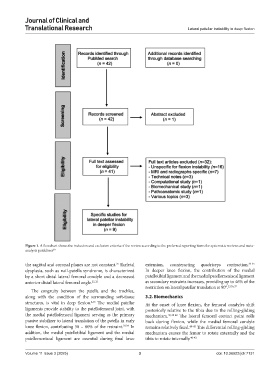
Figure 1. A flowchart shows the inclusion and exclusion criteria of the review according to the preferred reporting items for systematic reviews and meta-
analysis guidelines 20
the sagittal and coronal planes are not constant. Skeletal extension, counteracting quadriceps contraction. 33-35
25
dysplasia, such as nail-patella syndrome, is characterized In deeper knee flexion, the contribution of the medial
by a short distal lateral femoral condyle and a decreased patellotibial ligament and the medial patellomeniscal ligament
anterior distal lateral femoral angle. 25,32 as secondary restraints increases, providing up to 46% of the
restriction on lateral patellar translation at 90°. 33,36,37
The congruity between the patella and the trochlea,
along with the condition of the surrounding soft-tissue 3.2. Biomechanics
structures, is vital in deep flexion. The medial patellar At the onset of knee flexion, the femoral condyles shift
2,15
ligaments provide stability to the patellofemoral joint, with posteriorly relative to the tibia due to the rolling-gliding
the medial patellofemoral ligament serving as the primary mechanism. 30,38-40 The lateral femoral contact point rolls
passive stabilizer to lateral translation of the patella in early back during flexion, while the medial femoral condyle
knee flexion, contributing 50 – 60% of the restraint. 33,34 In remains relatively fixed. 40-42 This differential rolling-gliding
addition, the medial patellotibial ligament and the medial mechanism causes the femur to rotate externally and the
patellomeniscal ligament are essential during final knee tibia to rotate internally. 40-42
Volume 11 Issue 3 (2025) 3 doi: 10.36922/jctr.7131