
Page 14 - JCTR-11-3
P. 14
Journal of Clinical and
Translational Research Lateral patellar instability in deep flexion
Operative treatment for flexion instability should and subchondral quadriceps realignment) have been
address all documented etiological factors and typically recommended, particularly for treating congenital
involve a combination of procedures. 2,5,8,64-66 Two patellar dislocation. 4,64,67,68 A 4-in-1 quadricepsplasty,
types of surgical reconstruction are described: (i) soft- with or without lengthening, may be necessary in rare
tissue reconstruction, including lateral and medial cases with more severe forms of flexion instability to
approaches, 3,67,68 and (ii) soft–tissue reconstruction correct the externally rotated and shortened quadriceps
combined with bone reconstruction, such as elevation mechanism. 3,64,68 Z-lengthening of the quadriceps tendon
68
of the lateral femoral condyle. Contractures of the soft can be performed to restore its proper length. However,
2,4
tissues lateral to the patella are among the most important normal patellar tracking has also been reported without
2,64
contributing factors and must be addressed during surgery requiring quadriceps tendon lengthening.
(Table 4). 5,8,69 Therefore, lengthening and/or releasing all Biomechanically, the decreased resisting forces of the
involved structures is the first step in the reconstruction lateral femoral condyle are crucial in the reconstruction
process. process. 2,4,25 Any dysplastic shape of the lateral femoral
condyle, a pathologic terminal sulcus, and/or a false groove
The role of quadriceps tendon lengthening remains a must be corrected to restore the proper morphology
topic of discussion. Several quadricepsplasty techniques according to the proximal and distal spherical morphology
4,64
(i.e., sliding lengthening plasty of the lateral half of if present. This correction is determined by the condition
the distal quadriceps tendon, 4-in-1 quadricepsplasty, of the soft tissue after the release of contracted structures
and the patellar stability during higher flexion. There is
Table 3. Pathological factors for lateral patellar instability in a clear indication for additional bone reconstruction if
deep flexion patellar instability in flexion persists after extensive release
of the soft tissues.
Factors (most common in combination) 2‑5,7,25,64
Changing shapes of the lateral and medial condyle during knee flexion 3.5.1. Surgical technique
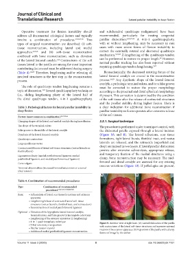
The shape of the terminal sulcus The procedure is performed under tourniquet control, with
False groove to the middle of the lateral condyle the dislocated patella exposed through a lateral incision
Dysplasia of the lateral femoral condyle (Figure 9A and B). The lateral adhesions, scar tissue
Quadriceps contracture formations, tight lateral bands, iliotibial tract, and vastus
Large quadriceps vector lateralis are released, and the retinacula (superficial and
Contractures/fibrosis of lateral soft tissue structures (vastus lateralis or deep) are incised in two layers. If lateral patellar dislocation
iliotibial band) persists after extensive adhesiolysis, appropriate release,
Ligamentous laxity (medial patellofemoral ligament, medial and temporary fixation of the medial structures using a
patellotibial ligament, and medial patellomeniscal ligament) clamp, bone reconstruction may be necessary. The mid-
Genu valgum femoral and distal condyle are assessed for any existing
osseous variations (Figure 10). If pathologies are present,
Torsional abnormalities (increased femoral antetorsion or external
tibial torsion)
A B
Table 4. Combination of recommended procedures
Type Combination of recommended
procedures 2‑5,8,33,35‑37,64,68,70,71
Basic • Adhesiolysis of lateral scar tissue formations and extensor
apparatus
• Lengthening/release of contracted lateral soft‑tissue
structures (vastus lateralis, iliotibial tract, and retinaculum)
• Reconstruction of medial patellofemoral ligament
Optional • Elevation of the hypoplastic lateral femoral condyle,
terminal sulcus, and false groove by incomplete osteotomy
• Lengthening of the extensor apparatus (Z‑lengthening)
• 4‑in‑1 quadricepsplasty technique Figure 9. Anterior view of right knee. (A) Lateral dislocation of the patella
• Tibial tuberosity transposition with contractures of the lateral soft-tissue structures and excessive external
• Patellar tendon transfer rotation of the extensor apparatus. (B) Reposition of the patella with a clamp.
• Additional medial patellotibial ligament reconstruction
Source of image by the author.
Volume 11 Issue 3 (2025) 8 doi: 10.36922/jctr.7131