
Page 13 - JCTR-11-3
P. 13
Journal of Clinical and
Translational Research Lateral patellar instability in deep flexion
A B A B
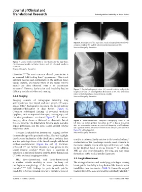
Figure 6. Radiographs of the right knee. Axial radiographs show (A) well-
centered patella at 30° and (B) lateral patellar dislocation at 60°.
Source of image by the author.
A B
Figure 5. Lateral patellar instability in deep flexion in the right knee.
(A) Dislocated patella in higher flexion and (B) relocated patella in
extension.
Source of image by the author.
dislocated. 2,3,5 The most common clinical presentation is
an unusual “odd-locking knee” appearance. Shortened
2,5
extensor muscles and contractures in the iliotibial band,
vastus lateralis, and lateral fibers of the rectus femoris
muscle are often observed. Pain is an uncommon
3
symptom. However, dysfunction and instability lead to Figure 7. Sagittal radiographs show (A) normal trochlea and patellar
difficulty in daily activities and running. height at 30° and (B) lateral patellar dislocation at 60°. The dotted line
refers to the flattened mid-femoral lateral condyle.
3.4.2. Imaging Source of image by the author.
Imaging consists of radiographs (standing long
anteroposterior, true lateral, and axial views), CT scans, A B
and/or MRI. Radiographs document the lateral patellar
2
subluxation/dislocation in deep flexion (Figure 6).
Common radiological findings of proximal trochlear
dysplasia, such as supratrochlear spur, crossing sign, and
2
trochlear prominence, are absent (Figure 7). In contrast,
imaging often shows a flattened or dysplastic lateral Figure 8. Three-dimensional computed tomography scans of the
femoral condyle. The distal lateral femoral angle may also left knee. (A) Lateral patellar dislocation in 60° of flexion (condition
appear pathologic, and the distal lateral femoral condyle after an unsuccessful attempt at proximal deepening trochleoplasty).
may be too short. (B) Dysplastic and flattened mid-femoral lateral condyle (same patient as
Figure 7A without patella).
CT scans (axial and three-dimensional imaging) confirm Source of image by the author.
the normal shape of the proximal trochlea. They also highlight
the decreased inclination of the distal lateral trochlear facet condyle at or immediately anterior to the terminal sulcus,
63
and the pathologic form of the lateral condyle with lateral contractures of the quadriceps muscle, most common in
subluxation/dislocation (Figure 8A and B). Contrast- the vastus lateralis muscle with signs of fibrosis, and rarely
enhanced CT can further visualize a false groove in the in the iliotibial band or rectus femoris. In addition,
3,63
lateral femoral condyle. When there is suspicion of MRI can also show elongation, thinning, and scar tissue
4
variations in the lateral femoral condyle, three-dimensional formations in the medial patellar ligaments.
CT is the preferred imaging modality.
3.5. Surgical treatment
MRI (two-dimensional and three-dimensional)
is another reliable modality to assess the bony and The etiological factors and underlying pathologies causing
cartilaginous morphology of the knee, particularly the lateral patellar instability in deep flexion differ from those for
trochlea. 22,61,62 MRI findings in patients with patellar instabilities close to extension (Table 3). Hence, the surgical
instability in flexion revealed injuries to the lateral femoral treatment is not the same and must be individually adapted. 2,5
Volume 11 Issue 3 (2025) 7 doi: 10.36922/jctr.7131