
Page 11 - JCTR-11-3
P. 11
Journal of Clinical and
Translational Research Lateral patellar instability in deep flexion
Figure 2. Axial view of the right knee. During extension, the lateral
trochlear facet (red arrow) is normally higher than the medial condyle. Figure 4. Lateral view of the left knee. Connections from the iliotibial
Source of image by the author. tract to the superficial lateral retinaculum, patellar tendon, patella, and
vastus lateralis tendon.
Source of image by the author.
contributing to patellar instability in deeper knee flexion.
The biomechanical sequence of patellar instability in deep
flexion is summarized in Table 2.
3.3. Common instability factors
Three factors are relevant for symptomatic patellar
instability: trochlear dysplasia, patella alta, and TT-TG
distance. 30
3.3.1. Trochlear dysplasia
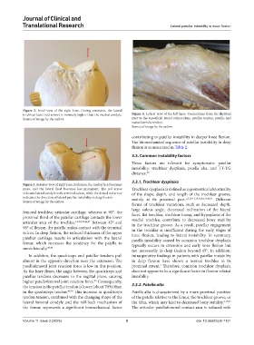
Figure 3. Anterior view of right knee. In flexion, the medial facet becomes
more, and the lateral facet becomes less prominent. The red arrow Trochlear dysplasia is defined as a geometrical abnormality
indicates lateral condyle with terminal sulcus, while the dotted red arrow of the shape, depth, and length of the trochlear groove,
indicates the direction of lateral patellar instability in deep flexion. mainly at its proximal part. 1,2,10-13,15,16,24,31,46,51 Different
Source of image by the author.
forms of trochlear variations, such as decreased depth,
large sulcus angle, decreased inclination of the lateral
femoral trochlear articular cartilage, whereas at 90°, the facet, flat trochlea, trochlear bump, and hypoplasia of the
proximal third of the patellar cartilage contacts the lower medial trochlea, contribute to decreased bony stability
articular area of the trochlea. 1,2,30,39,46,47 Between 45° and in the trochlear groove. As a result, patellar engagement
90° of flexion, the patella makes contact with the terminal in the trochlea is insufficient during the early stages of
sulcus. In deep flexion, the reduced thickness of the upper knee flexion, leading to lateral instability. In summary,
patellar cartilage results in articulation with the lateral patella instability caused by common trochlear dysplasia
femur, which increases the tendency for the patella to typically occurs in extension and early knee flexion but
move laterally. 46,48 not necessarily in deep flexion beyond 45°. In addition,
In addition, the quadriceps and patellar tendons pull intraoperative findings in patients with patellar instability
almost in the opposite direction near the extension. The in deep flexion have shown a normal trochlea in its
patellofemoral joint reaction force is low in this position. proximal extent. Therefore, common trochlear dysplasia
2
As the knee flexes, the angle between the quadriceps and does not appear to be a significant factor in flexion-related
patellar tendons decreases in the sagittal plane, causing instability.
higher patellofemoral joint reaction force. Consequently,
25
the tension in the patellar tendon is lower (about 70%) than 3.3.2. Patella alta
in the quadriceps tendon. 49,50 This increase in quadriceps Patella alta is characterized by a more proximal position
tendon tension, combined with the changing shape of the of the patella relative to the femur, the trochlear groove, or
lateral femoral condyle and the roll-back mechanism of the tibia, which may lead to decreased bony stability. 9,16,52
the femur, represents a significant biomechanical factor The articular patellofemoral contact area is reduced with
Volume 11 Issue 3 (2025) 5 doi: 10.36922/jctr.7131