
Page 33 - MI-1-1
P. 33
Microbes & Immunity The feature of bladder cancer stem cells
in response to different microenvironment conditions, et al. further establish a positive correlation between
allowing them to resist host defenses or treatment- tumor stemness and immunotherapy resistance or poor
induced damage. These adaptations are achieved through prognosis. The insights into BCSCs facilitate clinicians to
32
mechanisms including epigenetic modifications, epithelial- develop more effective treatment modalities and prognostic
mesenchymal transition (EMT), immune reprogramming, tools. This review provides a comprehensive overview of
and metabolic reprogramming. BCSC biomarkers, stemness maintenance, drug resistance,
In 1997, Bonnet and Dick from the University of Toronto metastasis, immune evasion, metabolic reprogramming,
in Canada successfully isolated TSCs for the first time from and targeted therapies.
acute myeloid leukemia. Recent research has demonstrated 2. Biology of normal bladder tissue
20
the presence of TSCs across various cancer types, including
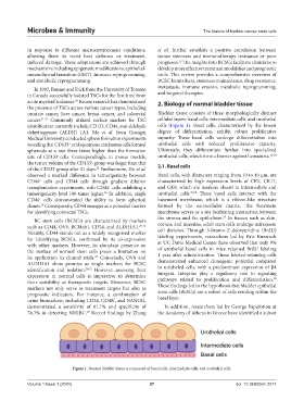
prostate cancer, liver cancer, breast cancer, and colorectal Bladder tissue consists of three morphologically distinct
cancer. 21-24 Commonly utilized surface markers for TSC cellular layers: basal cells, intermediate cells, and urothelial
identification currently include CD133, CD44, and aldehyde cells (Figure 1). Basal cells, characterized by the lowest
dehydrogenase (ALDH) 1A1. Ma et al. from Guangxi degree of differentiation, exhibit robust proliferative
Medical University conducted sphere formation experiments capacity. These basal cells undergo differentiation into
revealing that CD133 oral squamous carcinoma cells formed urothelial cells with reduced proliferative capacity.
+
spheroids at a rate three times higher than the formation Ultimately, they differentiate further into specialized
rate of CD133 cells. Correspondingly, in mouse models, urothelial cells, which form a barrier against hematuria. 33,34
-
the tumor volume of the CD133 group was larger than that 2.1. Basal cells
+
of the CD133 group after 21 days. Furthermore, Du et al.
25
-
observed a marked difference in tumorigenicity between Basal cells, with diameters ranging from 10 to 40 μm, are
CD44 cells and CD44 cells through gradient dilution characterized by high expression levels of CK5, CK17,
+
-
transplantation experiments, with CD44 cells exhibiting a and CK8, which are markers absent in intermediate and
+
tumorigenicity level 100 times higher. In addition, single urothelial cells. 35,36 These basal cells interact with the
26
CD44 cells demonstrated the ability to form spherical basement membrane, which is a ribbon-like structure
+
clones. Consequently, CD44 emerges as a potential marker formed by the extracellular matrix. The basement
26
for identifying colorectal TSCs. membrane serves as a site facilitating interaction between
37
the stroma and the epithelium. In tissues such as skin,
BC stem cells (BCSCs) are characterized by markers
such as CD44, OV6, BCMab1, CD24, and ALDH1A1. 27-31 cornea, and intestine, adult stem cells undergo infrequent
Notably, CD44 stands out as a widely recognized marker cell division. Through 5-bromo-2’-deoxyuridine (BrdU)
for identifying BCSCs, confirmed by its co-expression labeling experiments, researchers led by Eric Kurzrock
with other markers. However, its abundant presence on at UC Davis Medical Center have observed that only 9%
the surface of normal stem cells poses a limitation on of urothelial basal cells in mice retained BrdU labeling
its application in clinical trials. Conversely, OV6 and 1 year after administration. These labeled-retaining cells
27
ALDH1A1 show promise as single markers for BCSC demonstrated enhanced clonogenic potential compared
identification and isolation. 28,31 However, assessing their to unlabeled cells, with a predominant expression of β4
expression in normal cells is imperative to determine integrin. Integrins play a regulatory role in signaling
38
their suitability as therapeutic targets. Moreover, BCSC pathways related to proliferation and differentiation.
markers not only serve as treatment targets but also as These findings led to the hypothesis that bladder epithelial
prognostic indicators. For instance, a combination of stem cells (BESCs) are a subset of cells residing within the
urine biomarkers, including CD24, CD49f, and NANOG, basal layer.
demonstrated a sensitivity of 81.7% and specificity of In addition, researchers led by George Papafotiou at
74.3% in detecting NMIBC. Recent findings by Zhang the Academy of Athens in Greece have identified a subset
30
Figure 1. Normal bladder tissue is composed of basal cells, intermediate cells, and urothelial cells.
Volume 1 Issue 1 (2024) 27 doi: 10.36922/mi.2377