
Page 97 - MI-1-1
P. 97
Microbes & Immunity Dynamics between phage, bacteria, and mammalian cells
initiate the infection process. According to the findings in Overall, our results demonstrated that the dynamics
Figure 4D, the loss of sensitivity toward phage treatment between phages, bacteria, and mammalian cells can
was largely attributed to the bacteria’s capability to restrict affect the antibacterial efficiency of phages and the rate
the adsorption of phages onto the surface. This was of bacterial resistance development. While the findings
consistent with the findings reported by Barr et al. that highlighted the positive effects of mammalian cells toward
A. baumannii strains (AB900 and A9844) rapidly develop phage activity, a major limitation of the present study was
resistance against phage treatment (ΦFG02 and ΦCO01) the use of non-mucus-producing bronchial epithelial
via capsule loss, thereby disrupting phage adsorption. 43 BEAS-2B cells, which is an immortalized cell line. Phages
The interactions between bacteria and phages became and bacteria can behave differently with other cell types,
more complicated in the presence of epithelial cells. The such as mucus-producing and primary lung cells, which
previous studies have demonstrated that epithelial cells could affect their interactions. In addition, the simple
could also secrete chemokines and cytokines in response to three-component co-culture system could not completely
different stimuli to attract and stimulate the immune cells, mimic the complex in vivo environments. Hence, the
thereby aggravating inflammatory conditions. Schulz responses toward phage treatments may not have been
44
et al. also demonstrated that the lipopolysaccharide accurately represented in this study. For example, the
45
48
(LPS), a major component of the outer membrane of gram- generation of anti-phage neutralization antibodies
negative bacteria, could effectively stimulate the BEAS-2B and the modification of the microbiome during phage
49
and A549 epithelial cells to express immunoregulatory treatment may affect the effectiveness of phage therapy.
or inflammatory cytokines, such as soluble CD14, IL-6, Nonetheless, our findings revealed the remarkable
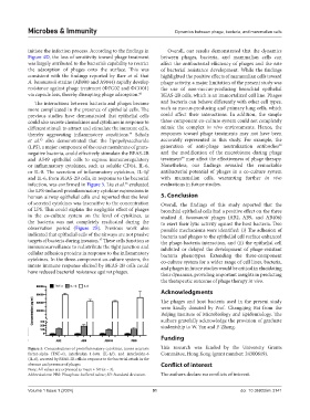
or IL-8. The secretion of inflammatory cytokines, IL-1β antibacterial potential of phages in a co-culture system
and IL-6, from BEAS-2B cells, in response to the bacterial with mammalian cells, warranting further in vivo
infection, was confirmed in Figure 5. Liu et al. evaluated evaluations in future studies.
46
the LPS-induced proinflammatory cytokine expressions in
human airway epithelial cells and reported that the level 5. Conclusion
of secreted cytokines was insensitive to the concentration Overall, the findings of this study reported that the
of LPS. This could explain the negligible effect of phages bronchial epithelial cells had a positive effect on the three
in the co-culture system on the level of cytokines, as studied A. baumannii phages (AB2, AB9, and AB406)
the bacteria was not completely eradicated during the to exert their lytic activity against the host bacteria. Two
observation period (Figure 2B). Previous work also possible mechanisms were identified: (i) The adhesion of
indicated that epithelial cells of the airways are not passive bacteria and phages to the epithelial cell surface enhanced
targets of bacteria during invasion. These cells function as the phage-bacteria interaction, and (ii) the epithelial cell
47
immunosurveillance to redistribute the tight junction and inhibited or delayed the development of phage-resistant
cellular adhesion proteins in response to the inflammatory bacteria phenotypes. Extending the three-component
cytokines. In the three-component co-culture system, the co-culture system for a wider range of cell lines, bacteria,
innate immune response elicited by BEAS-2B cells could and phages in future studies would be critical in elucidating
have reduced bacterial resistance against phages. their dynamics, providing important insights in predicting
the therapeutic outcome of phage therapy in vivo.
Acknowledgments
The phages and host bacteria used in the present study
were kindly donated by Prof. Changqing Bai from the
Beijing Institute of Microbiology and Epidemiology. The
authors gratefully acknowledge the provision of graduate
studentship to W. Yan and P. Zhang.
Funding
Figure 5. Concentrations of proinflammatory cytokines, tumor necrosis This research was funded by the University Grants
factor-alpha (TNF-α), interleukin 1-beta (IL-1β), and interleukin-6 Committee, Hong Kong (grant number: 24300619).
(IL-6), secreted by BEAS-2B cells in response to the bacterial attack in the
absence and presence of phages. Conflict of interest
Note: All values are expressed as mean ± SD (n = 3).
Abbreviations: PBS: Phosphate-buffered saline; SD: Standard deviation. The authors declare no conflicts of interest.
Volume 1 Issue 1 (2024) 91 doi: 10.36922/mi.3141