
Page 94 - MI-1-1
P. 94
Microbes & Immunity Dynamics between phage, bacteria, and mammalian cells
A B
C D
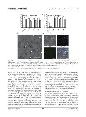
Figure 3. Bacteria and phage adsorption on the BEAS-2B cells in (A) a two-component system (bacteria/phage + BEAS-2B cells) and (B) a three-component
system (bacteria + phage + BEAS-2B cells). All values are expressed as mean ± SD (n = 3). (C) Giemsa staining of MDR-AB2 bacteria adhered onto the
BEAS-2B cells (bacteria clusters indicated with white arrows) Note: Scale bar: 100 μM; magnification: ×40. (D) Fluorescence images (×40 magnification)
of the adhesion of the FITC-labeled AB2 phages onto the BEAS-2B cell surface in a two-component system.
Note: Scale bars: 100 μM.
Abbreviations: DAPI: 4’,6-Diamidino-2-phenylindole; FITC: Fluorescein isothiocyanate; SD: Standard deviation.
bacteria lawn). According to Figure 4C, the resistance rate treated in the three-component system (41‒72% adsorption
of the phage-treated bacteria in the absence of BEAS-2B rate). The adsorption capability of AB2 and AB9 phages
cells was 100%, confirming that the regrowth of bacteria onto phage-treated bacteria in the absence of BEAS-2B
was accounted for by the phage-resistant phenotypes. In cells was completely diminished. The results suggested that
contrast, phage treatment in the presence of BEAS-2B the emergence of phage resistance was largely accounted
cells could significantly suppress resistance development for by the prevention of phage adsorption, thereby
toward phages. Among the three studied phages, AB2 and minimizing subsequent phage infections and leading to
AB406 phages could completely suppress the resistance bacterial regrowth. Overall, these results were consistent
development, and the AB9 phage could also efficiently with the bacteria-killing efficiency (Figure 2) that the AB2
reduce the resistance rate from 100% to 20% in the and AB406 phages have more prominent efficiency.
presence of epithelial cells. Bacteria recovered after phage
treatments, in the presence and absence of BEAB-2B cells, 3.4. Stimulation of cytokine secretion
were then subjected to phage adsorption assay. Figure 4D The inflammatory reaction of the BEAS-2B cells after
displays that phages incubated with PBS for 24 h were bacterial challenge in the absence and presence of phages
effectively adsorbed (100%) onto MDR-AB2 bacteria. was monitored after 24-h co-incubation by measuring the
However, the adsorption rate of phages onto bacteria with level of TNF-α, IL-1β, and IL-6. While negligible TNF-α
previous phage exposure significantly decreased, with was secreted, a moderate level of IL-1β and a high level of
the effect being more profound for bacteria treated in the IL-6 were produced by the epithelial cells in response to
two-component system (0‒23% adsorption rate) than that bacterial damage in the absence and presence of phages.
Volume 1 Issue 1 (2024) 88 doi: 10.36922/mi.3141