
Page 93 - MI-1-1
P. 93
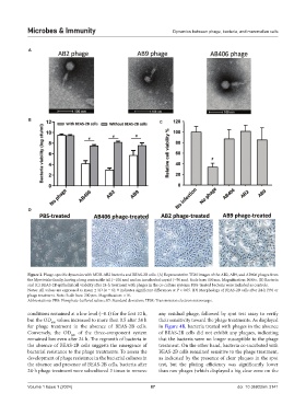
Microbes & Immunity                                     Dynamics between phage, bacteria, and mammalian cells


            A


            B                                                  C


            D


            Figure 2. Phage-specific dynamics with MDR-AB2 bacteria and BEAS-2B cells. (A) Representative TEM images of the AB2, AB9, and AB406 phages from
            the Myoviridae family, having a long contractile tail (~100 nm) and an icosahedral capsid (~50 nm). Scale bars: 100 nm. Magnification: 3000×. (B) Bacteria
            and (C) BEAS-2B epithelial cell viability after 24-h treatment with phages in the co-culture systems. PBS-treated bacteria were included as controls.
            Notes: All values are expressed as mean ± SD (n = 6); # indicates significant differences at P < 0.05. (D) Morphology of BEAS-2B cells after 24-h PBS or
            phage treatment. Note: Scale bars: 200 μm. Magnification: ×10.
            Abbreviations: PBS: Phosphate-buffered saline; SD: Standard deviation; TEM: Transmission electron microscope.

            conditions remained at a low level (~0.1) for the first 10 h,   any residual phage,  followed by spot  test  assay to verify
            but the OD  values increased to more than 0.5 after 24 h   their sensitivity toward the phage treatments. As displayed
                     600
            for phage treatment in the absence of BEAS-2B cells.   in Figure 4B, bacteria treated with phages in the absence
            Conversely, the OD  of the three-component system   of BEAS-2B cells did not exhibit any plaques, indicating
                             600
            remained low even after 24 h. The regrowth of bacteria in   that the bacteria were no longer susceptible to the phage
            the absence of BEAS-2B cells suggests the emergence of   treatment. On the other hand, bacteria co-incubated with
            bacterial resistance to the phage treatments. To assess the   BEAS-2B cells remained sensitive to the phage treatment,
            development of phage resistance in the bacterial cultures in   as indicated by the presence of clear plaques in the spot
            the absence and presence of BEAS-2B cells, bacteria after   test, but the plating efficiency was significantly lower
            24-h phage treatment were subcultured 3 times to remove   than raw phages (which displayed a big clear zone on the


            Volume 1 Issue 1 (2024)                         87                               doi: 10.36922/mi.3141