
Page 103 - MI-1-2
P. 103
Microbes & Immunity Klebsiella pneumoniae diagnosed by NGS
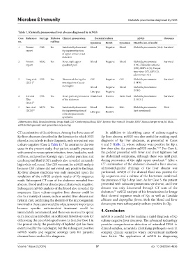
Table 1. Klebsiella pneumoniae liver abscess diagnosed by mNGS
Case Reference Sex/Age Diabetes Clinical presentation Bacterial culture mNGS Outcome
no. mellitus Specimen Result Specimen Microbe (no. of reads)
1 Present F/60 No Incidentally discovered Blood Negative Blood Klebsiella pneumoniae (144) Survived
report during investigation
of upper urinary tract
infection
2 Present M/82 Yes Fever, right upper Blood Negative Blood Klebsiella pneumoniae Survived
report quadrant pain (153), Klebsiella variicola
(256), HHV-6 (3), Torque
teno virus (37), EBV (1),
adenovirus D (1)
3 Zeng et al. F/59 Yes Discovered during the CSF Negative CSF Klebsiella pneumoniae Survived
(2021) 14 investigation of acute (13470)
meningitis Blood Negative Blood Klebsiella pneumoniae
Liver pus Negative (5318)
4 Xie and F/56 Yes Fever, pain on percussion Drain fluid Positive Drain fluid Klebsiella pneumoniae Survived
Zhu of the abdomen (119331)
(2021) 15
5 Luo et al. M/71 Yes Incidentally discovered Blood Positive BAL Klebsiella pneumoniae Died
(2023) 16 during investigation of Liver pus Positive (not mentioned)
subacute pneumonia
syndrome
Abbreviation: BAL: Bronchoalveolar lavage fluid; CSF: Cerebrospinal fluid; EBV: Epstein–Barr virus; F: Female; HHV: Human herpes virus; M: Male;
mNGS: Metagenomic next-generation sequencing.
CT examination of the abdomen. Among the three cases of In addition to identifying cases of culture-negative
Kp liver abscesses described in the literature in which NGS Kp liver abscess, mNGS was also useful for making rapid
played a crucial role in their diagnosis, only one patient was diagnosis of Kp liver abscesses, as presented in Cases
14
culture-negative (Case 3, Table 1). In contrast to the two 4 and 5 (Table 1), whose cultures were positive for Kp a
cases in the present study, that patient actually presented few days after the positive mNGS results. 15,16 For Case 4,
with central nervous system infection, fever, headache, neck the patient presented with fever and chest tightness but
stiffness, and positive Kernig’s sign. Lumbar puncture and no abdominal symptoms, although there was mild pain
15
cerebrospinal fluid (CSF) analysis also revealed extremely during percussion of the right upper quadrant. After a
high white cell count. The CSF was sent for mNGS analysis CT examination of the abdomen showed a liver abscess,
because CSF culture did not reveal any positive findings. ultrasound-guided drainage of the liver abscess was
Kp liver abscess syndrome was only suspected upon the performed. mNGS of the drained fluid was positive for
revelation of the mNGS analysis results of Kp sequence Kp sequences and a culture of the bacterium confirmed
reads. Subsequent CT scan of the abdomen revealed liver the presence of Kp 3 days later. As for Case 5, the patient
abscess. Blood and liver abscess pus cultures were negative. presented with subacute pneumonia syndrome, and liver
Subsequent mNGS analysis of the blood also revealed Kp abscess was only discovered through CT scan of the
16
sequences. Since culture-negative liver abscess could be abdomen. mNGS analysis of his bronchoalveolar lavage
due to a variety of causes, such as amebic liver abscess and fluid showed sequence reads of Kp, as well as Candida
hydatid cyst, confirming the identity of the microorganism albicans and Aspergillus flavus. Both the blood and liver
involved in these cases would be of paramount importance abscess pus were subsequently culture-positive for Kp.
because specific antimicrobial treatment could be
immediately commenced, and there was no need to spend 4. Conclusion
extra resources and effort on additional laboratory tests for mNGS is a useful tool for making a rapid diagnosis of Kp
delineating the microbiological cause. In fact, for Case 1 in culture-negative liver abscesses. The advanced technology
the present study, the possibility of hydatid cyst has been provides comprehensive detection of microbial DNA in
entertained by the radiologist, but the subsequent positive clinical samples, accurately identifying pathogens even in
mNGS results and negative serology tests for parasitic complex clinical scenarios where conventional methods
diseases have resolved the diagnosis. have failed. The application of mNGS in diagnosing
Volume 1 Issue 2 (2024) 97 doi: 10.36922/mi.4636