
Page 115 - MI-2-1
P. 115
Microbes & Immunity Anti-mouse CXCR5 monoclonal antibody
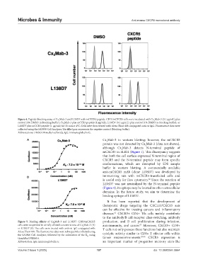
Figure 4. Peptide-blocking assay of Cx Mab-3 and L138D7 with mCXCR5 peptide. CHO/mCXCR5 cells were incubated with Cx Mab-3 (0.1 µg/mL) plus
5
5
control (1% DMSO in blocking buffer), Cx Mab-3 plus mCCR5 peptide (1 μg/mL), L138D7 (0.1 µg/mL) plus control (1% DMSO in blocking buffer), or
5
L138D7 plus mCCR5 peptide (1 μg/mL) for 30 min at 4°C. Cells were then treated with Alexa Fluor 488-conjugated anti-rat IgG. Fluorescence data were
collected using the SA3800 Cell Analyzer. The filled gray represents the negative control (blocking buffer).
Abbreviations: DMSO: Dimethyl sulfoxide; IgG: Immunoglobulin G.
A Cx Mab-3 in western blotting; however, the mCXCR5
5
protein was not detected by Cx Mab-3 (data not shown),
5
although Cx Mab-3 detects N-terminal peptide of
5
mCXCR5 in ELISA (Figure 1). This discrepancy suggests
that both the cell surface-expressed N-terminal region of
CXCR5 and the N-terminal peptide may form specific
conformations, which are disrupted by SDS sample
buffer in western blotting. A commercially available
anti-mCXCR5 mAb (clone L138D7) was developed by
B immunizing rats with mCXCR5-transfected cells and
is useful only for flow cytometry. Since the reaction of
60
L138D7 was not neutralized by the N-terminal peptide
(Figure 4), its epitope may be located in other extracellular
domains. In the future study, we aim to determine the
binding epitope of L138D7.
It has been reported that the development of
therapeutic drugs targeting the CXCL13/CXCR5 axis
can be effective for treating cancers and inflammatory
diseases. CXCR5+ CD4+ Tfh cells mainly contribute
61
to the antibody/B cell receptor class-switching, antibody
Figure 5. Binding affinity of Cx Mab-3 and L138D7. CHO/mCXCR5 production, and B cell proliferation during infection,
5
cells were suspended in serially diluted concentrations of Cx Mab-3 (A) autoimmunity, and cancer. Moreover, CXCR5+ CD8+
62
5
or L138D7 (B). The cells were treated with anti-rat IgG conjugated with T cells not only possess these functions but also maintain
Alexa Fluor 488. The fluorescence data were subsequently collected using cytolytic activity similar to CD8+ T effector cells within
the SA3800 Cell Analyzer, followed by the calculation of the K using
D
GraphPad PRISM 6. tumor microenvironments. 63-65 CXCR5 expression is
Abbreviation: IgG: Immunoglobulin G. an important marker of progenitor memory stem-like
Volume 2 Issue 1 (2025) 107 doi: 10.36922/mi.5664