
Page 173 - MI-2-3
P. 173
Microbes & Immunity Neurological and cranial involvement in SAPHO syndrome
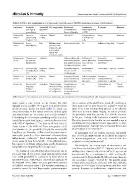
Table 1. Clinical and imaging features of previously reported cases of SAPHO syndrome associated with headache
Case report Headache Associated CSF composition Radiotracer Gadolinium- Treatment Response
type (age at symptoms accumulation in enhancing lesions
presentation) bone scintigraphy in MRI
DiMeco et al. Recurrent Allodynia, Normal First left rib Left parietal bone, Surgery Moderate
(2000) 14 incapacitating swelling extending to the
headaches (34) scalp
Hiwatani et al. Severe Fever Mild pleocytosis Sternoclavicular joints No Meloxicam 10 mg daily Complete
(2008) 15 headache (52) (101/mm , 98%
3
lymphocytes, 2%
polymorphs),
slightly high
protein (52 mg/dL)
Uematsu et al. Chronic severe Allodynia - Left parietal bone, left Left temporal Intravenous Moderate
(2012) 16 left temporal sterno-costo-clavicular muscle, left parietal methylprednisolone,
headache (50) joint, right femoral head, bone, and dura methotrexate
and intervertebral joints mater
Tsugawa et al. Recurrent right Allodynia, Normal Right bone, temporal, Frontotemporal Prednisolone 15 mg daily Complete
(2014) 2 frontal headache swelling sternum, and left muscle/fascia
(57) medial clavicula
Shiraishi et al. Severe forehead Swelling, Normal Frontoparietal bones, Right frontoparietal Intravenous Moderate
(2014) 17 headache (40) fever sternocostoclavicular bone marrow and methylprednisolone,
joint and sternum dura mater prednisolone 15 mg daily
Present case Recurrent left Arthralgias 20 leukocytes/µL, Frontal bones and No Sulphasalazine and Almost
hemicrania 44 erythrocytes/µL, peripheral joints, etoricoxib complete
headache 0.52 g/L glucose including the
(adolescence) 0.43 g/L proteins sternoclavicular and
sacroiliac
Abbreviations: SAPHO: Synovitis, acne, pustulosis, hyperostosis, and osteomyelitis, CSF: Cerebrospinal fluid; MRI: Magnetic resonance imaging.
skin, which is also present in this patient. The MRI due to osteitis of the skull bone, potentially resulting in
2
21
12
typically shows a pattern of T1 gadolinium enhancement brain infarction or even moyamoya disease. While no
in the involved tissues and bone (Table 1), which was signs of an active inflammatory process in the adjacent
not demonstrated in this patient where no contrast agent bone were present in brain imaging, we cannot preclude
was administered in the routine MRI initially ordered. the possibility that this process had already occurred
18
Considering the inflammatory cerebrospinal fluid, another in the past, leading to the formation of stenotic vessels.
possibility is aseptic meningitis, a condition also associated The other possibility is that the internal carotid artery is
15
with SAPHO syndrome. The absence of focal bone or constitutionally hypoplasic. Of note, hyperostosis in other
tissue lesions in the MRI and bone scintigraphy could segments (cervical) may lead to neurovascular lesions due
lend credence to this possibility. Despite the considerably to cervical spine compression. 22,23
long history of headaches in this patient, recurrent aseptic In agreement with the existing literature, our patient
meningitis would have been associated with underlying also showed an improvement of headache in response
inflammatory syndromes. Bone scintigraphy typically to immunosuppression (not only steroids but also
19
shows areas of focal hyperactivity in the affected bone, sulphasalazine and adalimumab). 2
but a pattern of diffuse enhancement on the frontal and
temporal bones has previously been reported. 17 We recognize that evident signs of osteomyelitis and
pustulosis, core features of SAPHO syndrome, were lacking
The finding of a stenotic internal carotid artery due to in this patient. However, the presence of focal hyperostosis
the narrowing of the carotid canal opening on the skull accompanied by a predominantly axial arthropathy
base could potentially be explained by hyperostosis of corroborates the diagnosis of SAPHO syndrome. Likewise,
the carotid canal. Narrowing of the proximal segments of the occasional vesicles reported by the patient could
the internal carotid artery has been previously reported correspond to acne and be associated with the syndrome,
in SAPHO syndrome cases. The vascular narrowing is although no overt signs of palmoplantar pustulosis were
20
probably caused by focal hypertrophic pachymeningitis present. Response to prednisolone, sulphasalazine, and
Volume 2 Issue 3 (2025) 165 doi: 10.36922/mi.4667