
Page 171 - MI-2-3
P. 171
Microbes & Immunity Neurological and cranial involvement in SAPHO syndrome
etiology. Besides, we review the existing evidence in the A B
literature to corroborate this association.
2. Case presentation
A 64-year-old female presented to the neurology
outpatient clinic in 2016 with complaints of headaches. She
described a heavyweight sensation on the left hemicrania
associated with periods of intense, sharp, frontal pain
that sometimes lasted for days, often interrupting sleep.
She had suffered from headaches during adolescence. C D
Over-the-counter anti-inflammatory drugs were seldom
effective in relieving her symptoms. Her past medical
history showed epidermoid papilloma of the tongue, which
was removed in 2014. Further inquiry of her past medical
history revealed inflammatory polyarthralgia occurring
in her hands, wrists, sternum, and lumbar spine. She also
reported occasional vesicles appearing in the lower back.
The neurological examination was unremarkable,
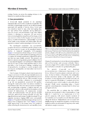
except for the discovery of allodynia in the left hemicrania. Figure 1. (A and B) Computed tomography-angiogram shows interruption
The patient reported no pain on the superficial temporal in the C4–C5 segments of the left internal carotid artery, after the carotid
arteries. A computed tomography (CT)-angiogram bulb, featuring a “pencil-tip pattern.” The left carotid canal is considerably
(Figure 1) demonstrated absent filling in the left internal narrower compared to the right one, due to hyperostosis of the walls.
carotid artery with a small carotid canal opening giving rise (C and D) Overall, the images suggest a hypoplasic and eventually dissected
to a narrow left internal carotid artery (shaped like a pencil left internal carotid artery. The posterior and anterior communicating
arteries granted collateral flow to its supraclinoid segment.
tip) that was more distally occluded. The posterior and
anterior communicating arteries granted collateral flow to
its supraclinoid segment. Her erythrocyte sedimentation Classical X-ray showed cervical and dorsal syndesmophytes
rate was 19 mm/s (normal level) and there were no other and calcified ischiatic and calcanean enthesitis. Human
anomalies in the blood panel findings, including levels leukocyte antigen (HLA) analysis showed that the patient
of autoantibodies. An attempt to control symptoms with does not harbor a risk allele for spondyloarthopaties.
escitalopram, amitriptyline, and alprazolam was found to The patient’s 45-year-old son developed
be ineffective. sternocostoclavicular inflammatory hyperostosis in his
Over the years, the headache attacks have become more forties, followed by palmo-plantar pustulosis and cystic
frequent and severe. In 2018, she reported daily severe and acne. He had no history of headaches. His scintigraphy
throbbing frontal headaches, which generally lasted several showed radiotracer uptake in the sternum and pubic
hours and impacted the left eye, with associated tearing symphysis. He was diagnosed with SAPHO syndrome and
and redness, a condition refractory to acetaminophen was on a treatment of interleukin-17 inhibitors, at the time
(1 gr tid) and prophylactic propranolol (80 mg/day). of writing this report, which resulted in a favorable clinical
During the examination, there was intense allodynia in outcome. Whole exome sequencing using a 34-gene
the left hemicrania. Given the atypical features of migraine panel for autoinflammatory diseases failed to detect any
and accompanying symptoms, a lumbar puncture was pathogenic variants.
performed to obtain cerebrospinal fluid for analysis, which
revealed 20 leukocytes/µL (13 mononuclear, normal: The possibility that our patient also had SAPHO
<5/µL), 44 erythrocytes/µL, 0.52 g/L glucose (normal: syndrome was considered. A trial of prednisolone 10 mg
<0.45 g/L), and 0.43 g/L proteins (normal: <0.5 g/L). No daily for 1 month led to an improvement of all the
oligoclonal bands were present. Results from brain CT and patient’s symptoms, with almost complete resolution of
magnetic resonance imaging (MRI) were unremarkable the headache, and it promptly recurred when she stopped
(Figure 2). The patient refused to undergo a temporal artery taking the medicine. Sulphasalazine and etoricoxib were
99
biopsy. Bone scintigraphy with mTC (Figure 3) showed initially enough to control the symptoms, but she further
slight hyperactivity in the frontal bones and peripheral needed escalation to adalimumab 40 mg every other week,
joints, including the sternoclavicular and sacroiliac joints. with almost complete remission.
Volume 2 Issue 3 (2025) 163 doi: 10.36922/mi.4667