
Page 56 - MI-2-3
P. 56
Microbes & Immunity Understanding lung development, health, and diseases
According to the prevailing historical theory, the lung
originated as an aquatic organ that resembles a swim
bladder. Due to the presence of epithelial cells lining the
2
swim bladder that contains surfactant proteins, lipids,
and lamellar bodies – organelles that transport surfactant
to the cell surface – sonic hedgehog expression in the
endoderm is essential for swim bladder development in
zebrafish. It is also known that the development of the
heart’s inflow and outflow tracts is regulated by signals
from the pulmonary and pharyngeal endoderm. This
implies that the cardiovascular system and lungs may have
coevolved. 3
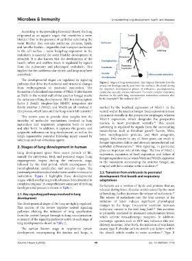
The developmental stages are regulated by signaling
pathways that drive the functional and structural changes Figure 1. Stages of lung development. This diagram illustrates how the
airway tree develops step-by-step from the trachea to the alveoli during
from embryogenesis to postnatal maturation. The the important developmental phases of embryonic, pseudoglandular,
formation of a localized expression of Nkx2-1 (also known canalicular, saccular, and alveolarization. To ensure complete respiratory
as Titf1) in the ventral wall of the anterior foregut marks function in the adult lung, microvascular maturation continues after
the initiation of lung development. SRY-box transcription birth. Copyright © The Author(s) 2017. 5
factor 2 (Sox2), wingless-type MMTC integration site
family member 2 (Wnt2), and Wnt2b are all involved in marked by the localized expression of Nkx2-1 in the
this process, which lasts until the lungs reach full maturity. 4 ventral wall of the anterior foregut. Sox2 expression is most
This review aims to provide clear insights into the prominent dorsally in the prospective esophagus, whereas
interplay of molecular mechanisms involved in lung Nkx2-1 expression, which designates the prospective
16
maturation and respiratory adaptation both in utero trachea, is most prominent ventrally. This spatial
and after birth. In addition, it captures the genetic and patterning is regulated by signals from the surrounding
epigenetic influences on lung development, as well as the mesenchyme, such as fibroblast growth factors, Wnts,
lung’s regenerative potential following injury caused by bone morphogenetic proteins, and their antagonist,
infectious and non-infectious agents. noggin. Deficiencies in any of these genes can result in
foregut separation failure and aberrant mesenchymal and
2. Stages of lung development in human epithelial differentiation. Wnt signaling, in particular,
17
plays an important role at this stage. The loss of Nkx2-1
Lung development spans three major periods of life, expression, expansion of Sox2 expression, and failure of
namely the embryonic, fetal, and postnatal stages. Lung foregut separation occur when Wnt2 and Wnt2b, expressed
organogenesis begins during the embryonic stage, in the mesoderm surrounding the anterior foregut, are
followed by the fetal period, which encompasses the coupled with beta-catenin in the endoderm. 18
pseudoglandular, canalicular, and saccular stages. The
postnatal period includes alveolarization and microvascular 2.2. Transition from embryonic to postnatal
maturation. Figure 1 highlights these developmental development: First breath and respiratory
stages, which overlap as growth advances from proximal to adaptations
peripheral regions. A comprehensive summary of the lung Surfactants are a mixture of lipids and proteins that are
5
developmental process is shown in Table 1.
released during labor. Alveolar stretch caused by the onset
19
2.1. Key signaling pathways involved in lung of breathing further increases the release of surfactants.
development The release of surfactants into the fetal airways at the
initiation of labor induces significant physiological
The developmental stages of the lung are tightly regulated. changes in the lungs. Concurrent secretion increases
This section of the review explores central signaling surfactant content in the fetal lung fluid. This secretion
20
pathways eliciting the initiation of lung specification is primarily mediated by increased catecholamine levels,
from the ventral foregut through to lung vascularization. which activate beta-adrenergic receptors. In addition,
summary of the signaling pathways active at each stage of purinergic agonists such as ATP may influence this pre-
lung development is shown in Table 2. delivery secretion. After birth, the initiation of ventilation
The earliest known stage in respiratory system causes type II alveolar cells to stretch and deform within
development, encompassing the trachea and lungs, is the alveoli, which results in more secretion. Type II
21
Volume 2 Issue 3 (2025) 48 doi: 10.36922/mi.7719