
Page 37 - OR-1-1
P. 37
is a promising strategy for developing and optimizing biological factors are included in the exosomes.
biomaterials with superior osteoinductive capacity. Especially, the SC-derived exosomes, as an important
114
New advancements also showed that the SCs respond to crosstalk vehicle between sensory neurons and osteocytes
external stimuli with biomaterial-mediated interventions, in the periosteum and Haversian systems, have been
like endogenous electric field formation, and express more used to improve the osteogenesis ability of MSCs while
neurogenic proteins to stimulate sensory nerve activity promoting vascularization and innervation within
118
and axonal outgrowth, further fostering vascularization the bone lesions (Figure 5). Moreover, it has been
and newborn bone development. 115-117 Given the critical demonstrated that the anti-inflammatory property of the
roles of the SCs within bone tissue development and SC-derived exosomes markedly facilitated macrophage
regeneration, a novel approach of loading the SCs and polarization toward the M2 phenotype rather than the
their derivatives into the bone biomaterials and the osteo- M1 phenotype, contributing to an anti-inflammatory
119
organoids has emerged as an innovative strategy to repair microenvironment for osteocyte growth. Therefore,
the refractory bone defects. the exosomes are usually engineered into a sustained-
release agent, in which the neurotrophic factors stored
Exosomes, also known as extracellular vesicles from in the exosomes can be released from biomaterials to
the paracrine pathway, are considered a promising stimulate regeneration of the innervated bone tissues.
therapy for bone regeneration because abundant Apart from the SCs, MSCs also produce exosomes with
A B
C
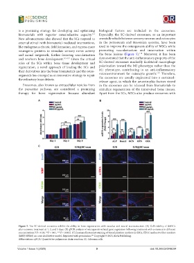
Figure 5. The SC-derived exosomes exhibit the ability in bone regeneration with vascular and neural reconstruction. (A) Cell viability of BMSCs
after exosome treatment at 1, 3, and 5 days. (B) qPCR analysis of osteogenesis-related gene expression following treatment with exosomes in different
concentrations (*P < 0.05, **P < 0.01, ***P < 0.001). (C) Immunofluorescent staining of vascularization markers (a-SMA, CD31) and nerve fiber markers
(MBP, NF200) on a rat skull defect model. Reprinted with permission. Copyright © 2023, KeAi Publishing.
121
Abbreviations: qPCR: Quantitative polymerase chain reaction; SC: Schwann cells.
Volume 1 Issue 1 (2025) 9 doi: 10.36922/OR8294