
Page 135 - OR-1-2
P. 135
A
B
C
D
E
F G H
I J
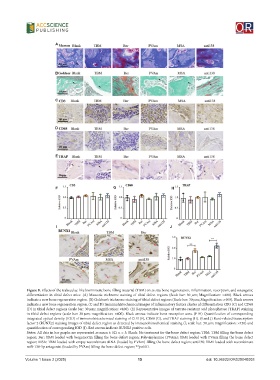
Figure 8. Effects of the trabeculae-like biomimetic bone-filling material (TBM) on in situ bone regeneration, inflammation, resorption, and osteogenic
differentiation in tibial defect mice. (A) Masson’s trichrome staining of tibial defect regions (Scale bar: 30 μm; Magnification: ×400). Black arrows
indicate a new bone regeneration region. (B) Goldner’s trichrome staining of tibial defect regions (Scale bar: 30 μm; Magnification: ×400). Black arrows
indicate a new bone regeneration region. (C and D) Immunohistochemical images of inflammatory factors cluster of differentiation CD3 (C) and CD68
(D) in tibial defect regions (scale bar: 30 μm; magnification: ×400). (E) Representative images of tartrate-resistant acid phosphatase (TRAP) staining
in tibial defect regions (scale bar: 30 μm; magnification: ×400). Black arrows indicate bone resorption area. (F-H) Quantification of corresponding
integrated optical density (IOD) of immunohistochemical staining of CD3 (F), CD68 (G), and TRAP staining (H). (I and J) Runt-related transcription
factor 2 (RUNX2) staining images of tibial defect region as detected by immunohistochemical staining (I; scale bar: 30 μm; magnification: ×450) and
quantification of corresponding IOD (J). Red arrows indicate RUNX2 positive cells.
Notes: All data in bar graphs are represented as mean ± SD, n = 3; Blank: No treatment for the bone defect region; TBM: TBM filling the bone defect
region; Ber: TBM loaded with bergamottin filling the bone defect region; Polyvinylamine (PVAm): TBM loaded with PVAm filling the bone defect
region; MSA: TBM loaded with empty recombinant tRNA (loaded by PVAm) filling the bone defect region; anti138: TBM loaded with recombinant
miR-138-5p antagonist (loaded by PVAm) filling the bone defect region; **p<0.01.
Volume 1 Issue 2 (2025) 15 doi: 10.36922/OR025040003