
Page 72 - TD-2-2
P. 72
Tumor Discovery SRT with SIP planning for synovial sarcoma
®
performed. Celiac axis and common hepatic artery were a 4D was acquired thanks to a respiratory belt (Philips );
clearly infiltrated by tumor and a surgical clip was left for second, a triphasic contrast-enhanced CT study was
post-operative guidance purposes. Pathologic examination performed. Both the studies used a slice thickness of
confirmed an SS relapse, with a vital cellularity of 40%, and 1.5 mm.
also confirmed the specimen translocation of locus SS18 Planning was performed on average CT to take into
(SYT-18q11.2). All four lymph nodes removed resulted account tumor and organs at risk (OARs) motion. In
negative for tumor involvement. Moreover, pathologic details, on Velocity (Varian ) treatment planning system
®
report confirmed the presence of invasive tumor at surgical (TPS), each of the ten respiratory phases was exploited to
margin (extensively R2 resection). The patient recovered define the position of the tumor, the stomach, duodenum,
without major complication and was discharged in good jejunum, and colon.
clinical conditions; acute toxicity was represented by
limitation of solid oral intake determined by G2 dyspepsia As a result, ten regions of interest were created for
in the following 2 months. the stomach, duodenum, and the critical area of jejunum
proximal to target. Moreover, internal margin (IM)
Taking into account the macroscopic persistence of representing the maximum position of the stomach,
disease, at the end of June 2021, the patient was evaluated by duodenum, and jejunum was created. For stomach and
a radiation oncologist that proposed a normofractionated jejunum near to the target, a planning respect volume
treatment consisting of 59.4 Gy in 33 fractions versus (PRV) was crated with an isotropic expansion of 2 mm.
60 Gy in 30 fractions on the sites of persistent disease.
A second approach was performed at our center, with the For target definition, exploiting all the information
propose to study the feasibility of an hypofractionated derived from diagnostic CT and contrast-enhanced planning
schedule, optimally stereotactic body RT in 5 – 6 fractions. CT, after co-registration to each respiratory phase, a gross
Our choice for hypofractionation was aimed at maximum tumor volume (GTV) was defined on each respiratory
tumor control probability, given the well-known intrinsic phase. The sum of GTV of each phase formed the internal
radioresistance of the histologic subtype to standard target volume (ITV) that virtually represents the area, in
normofractionated regimens. which GTV moves during the respiratory cycle. A margin
accounting for set-up errors and unpredictable organ motion
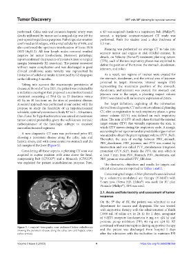
A new diagnostic CT scan was performed prior RT, was added to obtain the planning target volume (PTV_Full).
showing a persistent disease along the celiac axis and Thereafter, the area of overlap between PRV_stomach,
hepatic artery, and with tissue contact to stomach and the PRV_duodenum, PRV_jejunum, and PTV was created by
left margin of the liver (Figure 5).
intersection and was called PTV_simultaneous integrated
Considering all these aspects, a planning CT scan was protection (PTV_SIP). Inside the PTV, the area which is
acquired in supine position with arms above the head, at least 7 mm from PRV_Stomach, PRV_duodenum, and
®
®
compressing Belt (CIVCO ) and a Monarch (CIVCO ) PRV_jejunum was called PTV_SIB dose.
was exploited for patient immobilization purpose. First, The dosimetric objectives and results for targets and
critical structures are reported in Tables 1 and 2.
Concerning technique, 6 Mev photons beams delivered
by a volumetric-modulated arc therapy (V-MAT) with
5-mm jaws (Versa HD, Elekta ) was used; for RT plan
®
Pinnacle (Philips ), TPS was used.
®
2.1. Acute and late toxicity and assessment of tumor
response
th
On the 5 day of RT, the patient was admitted to our
department for nausea and dyspepsia. She was treated
with supportive therapy with the administration of fluids
(1000 mL of saline e.v. in 24 h) for 2 days, antagonist
of 5-HT3 receptors (ondansetron 8 mg e.v. q24 h) and
protonic pump inhibitors (PPI; 40 mg e.v. q24 h). RT
continued without interruption during supportive therapy
Figure 5. Computed tomography scan performed before radiotherapy
showing the persistent disease along the celiac axis and hepatic artery and the patient was discharged from hospital 5 days
(white arrows). after the admission with the indication to maintain PPI
Volume 2 Issue 2 (2023) 4 https://doi.org/10.36922/td.356