
Page 70 - TD-2-2
P. 70
Tumor Discovery SRT with SIP planning for synovial sarcoma
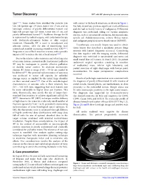
type [11-15] . Some studies have stratified the patients into with contact to the hearth structures, as shown in Figure 1.
low-risk (patient age <25 years, tumor size <5 cm, and no The lady denied any previous significant medical illnesses
histologic evidence of poorly differentiated tumor) and and she had no family history of malignancy. Differential
high-risk groups (age ≥25 years, tumor size ≥5 cm, and diagnosis was performed, ruling out various neoplastic
poorly differentiated tumor) . Backbone therapy for SS entities, such as sarcomatoid carcinoma, leiomyosarcoma,
[11]
is represented by radical surgery, with polychemotherapy spindle cell rhabdomyosarcoma, solitary fibrous tumor,
with ifosfamide-adriamycin before or after surgical and malignant peripheral nerve sheath tumor (MPNST).
treatment. Classically, RT is used in neoadjuvant and
adjuvant context, with the aim of maximizing local Transthoracic core-needle biopsy was used to obtain
control and possibly increasing overall survival (OS) [16,17] . tissue biopsy that described a neoplastic process (huge
Prognosis in case of Rx/1 resection is worse, with a possible necrosis with limited diagnostic accuracy). Considering
impact of RT to reduce the risk of local relapse [18-20] . this data together with the imaging results, differential
diagnosis was restricted to sarcomatoid carcinoma and
Due to the relative radio-resistance and often large size small round blue cell tumors. In March 2015, the patient
of sarcoma lesions, conventionally fractionated palliative underwent surgical operation consisting in resection
RT may be inadequate to provide effective palliation of mediastinal mass, inferior right lobectomy, and
or durable tumor control. To eradicate microscopic partial resection of right diaphragm with bovine patch
disease, RT doses at range of 60 – 70 Gy are needed to reconstruction. No major perioperative complications
[21]
be delivered . The potential radioresistance of sarcomas occurred.
was attributed to tumor cell capacity for sublethal
damage repair, as implied by the initial large shoulder Results of pathologic examination were consistent with
on their survival curve [22,23] . One of the radiobiological the diagnosis of poorly differentiated SS with invasion of
characteristics of sarcoma cells is their relatively low costal muscle, visceral pleura, and mediastinum, in close
(0.5 – 5.4) α/β ratio, suggesting that such tumors may proximity to the pericardial sierosa. Margin status was
be more vulnerable to higher dose per fraction. This R1 with microscopic positivity in the right costal muscle.
ratio, theoretically, may justify the use of larger-than- The diagnosis was supported by immunochemistry
standard fractionation to achieve significant cell-kill by that showed positivity for Bcl2 and negativity for CD34,
RT . Stereotactic RT (SRT) technique enables delivery CD99, Ckpan S100, and SOX10. The molecular profile of
[24]
of high dose to the tumor in a relatively small number of disease showed translocation of locus SS18 (SYT-18q11.2).
fractions (generally from 1 to 8), potentially overcoming Figure 2A and B show histologic image and positive stain
radioresistance of some histological cancer subtypes. In for Bcl2.
SRT, the therapeutic ratio is optimized through delivery
of highly conformal dose distributions with steep dose All nodes analyzed were negative for disease
fall-off with the aim of optimal absorbed dose in the dissemination. The patient progressively recovered
target volume combined with minimal normal-tissue
irradiation. Despite these considerations, the impact of
RT in the context of unresectable or macroscopically
positive resection is poorly understood and classically
considered for palliative intent. The relevance of our case
report is manifold. Our analysis applies cutting-edge
technique together with innovative planning technique
to maximize therapeutic index and to minimize side
effects. Our considerations involve the analysis of the
choices on the clinical and radiobiological point of view,
contributing to enrich the evolving literature in the field.
2. Case presentation
In August 2014, a 36-year-old lady experienced worsening
of dyspnea and upper back pain after childbirth. In
November 2014, a thorax and abdomen computed
tomography (CT) scan with and without contrast agent was Figure 1. Thorax and abdomen computed tomography scan showing a
huge thoracic/posterior mediastinum right mass infiltrating lung and
performed on her, which showed a huge thoracic/posterior diaphragm and with contact to the hearth and esophageal structures
mediastinum right mass infiltrating lung, diaphragm and (white arrows).
Volume 2 Issue 2 (2023) 2 https://doi.org/10.36922/td.356