
Page 32 - TD-3-2
P. 32
Tumor Discovery Immunophenotypic patterns of childhood acute leukemia
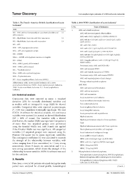
Table 1: The French–America–British classification of acute Table 2: 2016 WHO classification of acute leukemia 9
leukemia 8
S. No. Types of leukemias
AML ALL 1. AML and related neoplasms
M0 – AML with no Romanowsky or cytochemical evidence of L1 AML with recurrent genetic abnormalities
differentiation
AML with t (8;21) (q22;q22.1); RUNX1‑RUNX1T1
M1 – Myeloblastic leukemia with little maturation L2
AML with in v (16) (p13.1q22) or t (16;16) (p13.1;q22);
M2 – Myeloblastic leukemia with maturation L3 CBFB‑MYH11
M3 – APL APL with PML‑RARA
M3h – APL, hypergranular variant AML with t (9;11) (p21.3;q23.3); MLLT3‑KMT2A
M3v – APL, microgranular variant AML with t (6;9) (p23;q34.1); DEK‑NUP214
M4 – AMML AML with inv (3) (q21.3q26.2) or t (3;3) (q21.3;q26.2);
M4eo – AMML with dysplastic marrow eosinophils GATA2, MECOM
M5 – AMoL AML (megakaryoblastic) with t (1;22) (p13.3;q13.3);
RBM15‑MKL1
M5a – AMoL, poorly differentiated
M5b – AMoL, differentiated Provisional entity: AML with BCR‑ABL1
AML with mutated NPM1
M6 – “Erythroleukemia”
AML with biallelic mutations of CEBPA
M6a – AML with erythroid dysplasia
Provisional entity: AML with mutated RUNX1
M6b – Erythroleukemia
AML with myelodysplasia-related changes
M7 – Acute megakaryoblastic leukemia (AMkL)
Therapy-related myeloid neoplasms
Abbreviations: AML: Acute myeloid leukemia; APL: Acute
promyelocytic leukemia; AMML: Acute myelomonocytic leukemia; AML NOS
AMol: Acute monoblastic leukemia; ALL: Acute lymphoblastic AML with minimal differentiation
leukemia.
AML without maturation
2.4. Statistical analysis AML with maturation
Continuous data were reported as mean ± standard Acute myelomonocytic leukemia
deviation (SD) for normally distributed variables and Acute monoblastic/monocytic leukemia
as median with an interquartile range (IQR) for skewed Pure erythroid leukemia
variables. Categorical data were reported as percentages. Acute megakaryoblastic leukemia
P < 0.05 was considered statistically significant. We used Acute basophilic leukemia
STATA 14 software for statistical analysis. All continuous Acute panmyelosis with myelofibrosis
variables were assessed for normal or skewed distribution Myeloid sarcoma
(SD > 40% of mean). For variables with a skewed
distribution, the median (IQR) was reported. Comparisons Myeloid proliferations related to Down syndrome
of more than two unpaired groups were performed TAM
using the Kruskal–Wallis test, with Dunn’s test applied Myeloid leukemia associated with Down syndrome
if the Kruskal–Wallis test was significant. All categorical Blastic plasmacytoid dendritic cell neoplasm
variables (>2 unpaired groups) were assessed using the Acute leukemias of ambiguous lineage
Pearson Chi-square test to assess significant variability. Acute undifferentiated leukemia
The contingency coefficient (C) was calculated to measure MPAL with t (9;22)(q34.1;q11.2); BCR‑ABL1
the strength of the relationship between variables, with
values ranging from 0 (no association) to 1 (very strong MPAL with t (v; 11q23.3); KMT2A rearranged
association), where 0 means no association and 1 is a MPAL, B/myeloid, NOS
very strong association, which shows the strength of the MPAL, T/myeloid, NOS
relationship between variables. SATA 14 software was used 2. B-lymphoblastic leukemia/lymphoma
for data analysis. B-lymphoblastic leukemia/lymphoma, NOS B-lymphoblastic
leukemia/lymphoma with recurrent genetic abnormalities
3. Results B-lymphoblastic leukemia/lymphoma with t (9;22)
Data from a total of 68 patients obtained during the study (q34.1;q11.2); BCR‑ABL1
period were analyzed for clinical profile, hematological (Cont’d...)
Volume 3 Issue 2 (2024) 3 doi: 10.36922/td.2545