
Page 20 - TD-3-4
P. 20
Tumor Discovery Expert consensus of NUT carcinoma
presents as rapidly growing masses, often associated with patients may exhibit early, moderate osteolytic metastases
pain and low-grade fever. Between 60% and 77% of cases without obvious clinical symptoms. Metastases may not
exhibit distant metastasis at the initial diagnosis, 17,18,30-32 be present initially but can develop during treatment. It is
with approximately 50.9% of patients in our dataset recommended that a comprehensive, whole-body contrast-
classified as stage IV. The most common sites of distant enhanced CT scan of the chest, abdomen, pelvis, and brain
metastasis include the bones, lungs, pleura, liver, brain, MRI is performed at the initial visit. If feasible, PET-CT or
adrenal glands, kidneys, and skin. In addition, 68% of a whole-body bone scan should be performed to establish
cases show involvement of regional lymph nodes. NUT baseline data for subsequent treatment planning and
31
carcinoma typically originates along the midline, with efficacy assessment.
approximately 51% of cases occurring in the thoracic/ Imaging findings in NUT carcinoma are typically
mediastinal region. In our data, 45.4% of cases were of nonspecific, resembling those of other common malignant
33
thoracic origin, and 31.2% were of head-and-neck origin. solid tumors in the same anatomical location. These
Clinical manifestations of thoracic NUT carcinoma are findings are characterized by non-uniform enhancement
often dominated by a persistent cough, with other common of low-density masses with significant invasiveness.
36
symptoms including wheezing, chest tightness, dyspnea, Primary lung NUT carcinoma typically presents as
thoracic pain, shoulder pain, back pain, hemoptysis or centrally located masses with invasive, infiltrative
bloody sputum, and fever. 34,35 Beyond the thoracic region, growth patterns, and irregular shapes. About 36.8% of
NUT carcinoma frequently involves the head and neck, cases exhibit pleural effusion, and 57.9% are associated
particularly the sinonasal area. Most head-and-neck with obstructive atelectasis or obstructive pneumonia.
31
37
NUT carcinoma cases originate in the nasal cavity, with Head-and-neck NUT carcinoma typically manifests as
30.2% involving the paranasal sinuses and 14.3% affecting large, poorly defined, and space-occupying masses with
the salivary glands. Clinical manifestations of head-and- internal necrosis and hemorrhage. These tumors often
neck NUT carcinoma include pain at the lesion site, firm
swelling of the skin, difficulty opening the mouth, nasal
congestion, and other symptoms. At the time of diagnosis,
56.7% of head-and-neck NUT carcinoma patients show
invasion of surrounding tissues, with 26.7% exhibiting
regional lymph node involvement. Patients with head-
12
and-neck NUT carcinoma may also exhibit pain, swelling,
decreased vision, diplopia, facial numbness, choking
on liquids, epistaxis, and other symptoms due to tumor
invasion of adjacent tissues.
4.3. Imaging manifestations
Computed tomography (CT), magnetic resonance imaging
(MRI), ultrasound, positron emission tomography
(PET)-CT, and other imaging modalities are crucial
for the diagnosis, clinical staging, treatment response
assessment, and follow-up monitoring of NUT carcinoma.
Endoscopic examinations may also be performed when
appropriate, depending on the anatomical sites involved.
Contrast-enhanced CT or MRI is recommended for
anatomical regions affected by NUT carcinoma. Due to the
high malignancy of NUT carcinoma, distant metastasis
can occur early, with metastases commonly observed
in multiple organs. The skeleton is the most frequent
metastatic site, although metastases can occur in other
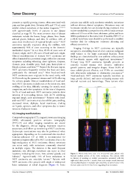
locations, including the brain, liver, adrenal glands, and Figure 3. Whole-body bone scan of a 30-year-old male patient with
kidneys. Approximately 75% of bone metastases in thoracic nuclear protein of the testis carcinoma. The patient, with
31
NUT carcinoma are osteolytic lesions. Patients with no history of smoking or alcohol consumption, presented with the
31
right-sided chest pain for 1 month. Bone scan images reveal abnormal
NUT carcinoma often present with single or widespread radionuclide shadows in multiple right-sided ribs, vertebral bodies in the
bone metastases at initial diagnosis (Figure 3). Some thoracic spine and the left iliac bone, suggesting bone metastasis.
Volume 3 Issue 4 (2024) 12 doi: 10.36922/td.4904