
Page 21 - TD-3-4
P. 21
Tumor Discovery Expert consensus of NUT carcinoma
invade adjacent tissues, such as the sinus walls, muscles, NGS, and liquid biopsy for molecular testing. However,
and nerves, and may involve cervical lymph nodes. On samples for molecular testing must undergo enrichment
12
CT imaging, head-and-neck NUT carcinoma typically and quality control to ensure reliability. It is essential to
appears as low-density masses with internal necrosis and note that tissue samples obtained through endoscopy,
hemorrhage. fine-needle aspiration, or core-needle biopsy may not fully
represent the morphology and structure of the tumor.
In contrast, MRI findings of NUT carcinoma typically
show low-signal intensity on T1-weighted images and This limitation could hinder the identification of typical
histological features, such as abrupt keratinization in NUT
high-signal intensity on T2-weighted images, along carcinoma, thereby increasing the diagnostic challenge.
41
36
with heterogeneous enhancement. Both primary and Since NUT carcinoma is frequently diagnosed in advanced
metastatic lesions in thoracic and head-and-neck NUT stages when tumor samples cannot be obtained through
carcinoma exhibit high fluorodeoxyglucose uptake on surgery, liquid biopsy samples such as circulating-tumor
PET-CT imaging. Lung NUT carcinoma tumors typically DNA (ctDNA) and high-throughput sequencing can be
31
have a maximum standardized uptake value (SUV) considered supplementary diagnostic tools. In addition,
exceeding 10, with an average SUV of 12, ranging from 5 endoscopy, fine-needle aspiration, or core-needle biopsy
to 40. Extra-pulmonary NUT carcinoma tumors have an may still be helpful in these cases.
average SUV of 13.8, ranging from 4.5 to 64.1. 38,39 Imaging
features of NUT carcinoma in other locations typically 4.4.2. Pathological histological features
include primary masses accompanied by metastatic lymph Due to its rarity and nonspecific manifestations, NUT
node enlargement and widespread distant metastases. carcinoma is often misdiagnosed during the initial
These features resemble those of advanced tumors in the pathological examination. In most cases, NUT carcinoma
respective sites, posing challenges in distinguishing from lacks specific histological features, with cancer cells
other malignancies. 38,39 While bone metastases can be typically showing poorly differentiated morphology.
42
evaluated using bone scintigraphy or PET-CT, current Approximately 33% to 40% of NUT carcinomas may exhibit
data suggest that NUT carcinoma bone metastases are focal squamous differentiation with abrupt keratinization
predominantly osteolytic. Since bone scintigraphy relies (Figure 4). In such cases, squamous cells can abruptly
on enhanced sodium phosphate metabolism driven by form small, round cell clusters with abundant keratinized
osteoblastic activity, PET-CT is recommended for a more cytoplasm and nuclear shrinkage, lacking stratification, and
40
accurate assessment. gradual differentiation processes. The stroma may show
43
4.3.1. Recommendation 1
A
NUT carcinoma frequently presents as large masses,
often accompanied by regional lymph node metastases
and distant metastases. Due to its lack of specificity, its
imaging features closely resemble those of advanced
tumors in the corresponding anatomical regions. Bone
metastases are commonly observed in NUT carcinoma,
and clinicians should be particularly vigilant when patients B
present with extensive bone metastases at the time of
initial diagnosis. Enhanced CT/MRI examinations of the
relevant anatomical sites and bone scans or PET-CT (more
recommended) are beneficial for staging and treatment
evaluation (level of evidence: Grade 4; recommendation
level: Strong recommendation).
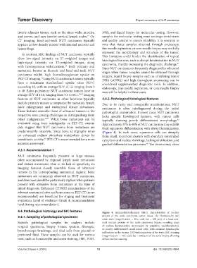
4.4. Pathological histology and IHC features Figure 4. Immunohistochemical (IHC) characteristics of nuclear
4.4.1. Sampling of pathological specimens protein of the testis carcinoma tumor tissue. (A) Hematoxylin and
eosin stain (magnification = 10×; scale bar = 200 µm) of a head-and-
Suitable pathological samples for analysis include neck nuclear protein of the testis carcinoma biopsy, revealing areas
surgical specimens, biopsy tissues, sputum, fiberoptic of sudden keratinization surrounded by primitive, undifferentiated,
bronchoscope brushings, and shed cells from pleural or or poorly differentiated small round cells, with minimal lymphocytic
infiltration in the stroma. (B) Nuclear protein of the testis IHC staining
peritoneal fluid. These samples can be used for various (magnification = 10×; scale bar = 200 µm) of the same biopsy, showing
tests, such as hematoxylin and eosin staining, IHC, FISH, diffuse nuclear staining.
Volume 3 Issue 4 (2024) 13 doi: 10.36922/td.4904