
Page 110 - TD-4-1
P. 110
Tumor Discovery HHT inhibits pancreatic cancer progress
kit (Beyotime, China) according to the manufacturer’s One-way analysis of variance (ANOVA) was used for
instructions. Luminescence intensity was measured using comparisons among three or more groups defined by
a microplate reader (Thermofisher Scientific, USA) and a single factor, whereas two-way ANOVA was used for
normalized to the protein content of the cell lysates. comparisons involving two factors. The least significant
difference post hoc test was conducted following ANOVA
2.12. NAD /NADH measurement to compare group means. A P < 0.05 was considered
+
PANC-1 cells were treated with HHT at 50 nM and 100 nM statistically significant.
for 24 or 48 h. Intracellular total NAD (NAD total ) and
NADH content were measured using a commercial assay 3. Results
kit (Beyotime, China) according to the manufacturer’s 3.1. HHT inhibited the growth and proliferation of
protocol. The NAD /NADH ratio was calculated according PDAC cells in vitro
+
to the following formula.
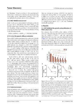
The cytotoxic effect of HHT on the viability of PDAC
[NAD ]/[NADH] = ([NAD total ] − [NADH])/[NADH] cells was evaluated using the CCK-8 assay. It was shown
+
that the viability of PANC-1 cells was significantly
2.13. In vivo therapeutic efficacy evaluation
inhibited by HHT in a dose- and time-dependent manner
First, an HHT toxicity assessment was conducted in healthy (Figure 1A). The IC50 value of HHT for PANC-1 at 24 hs
mice to determine a safe dosage for therapeutic evaluation. was 23.11 nM. Similarly, the IC50 values for SW1990 cells
Briefly, healthy female C57BL/6 mice (6 – 8 weeks old, and Pan02-mCherry cells at 24 h were 144.00 nM and
~20 g) were randomly divided into five groups (n = 3). 20.67 nM, respectively (Table S1). The effect of HHT on the
®
HHT solution was prepared in 0.4% (w/v) Soluplus proliferation of PANC-1 cells was examined using the CFSE
(BASF SE, Germany) in PBS, with four dosages including
0.5, 1, 2, and 4 mg/kg. A PBS solution containing 0.4% A
®
Soluplus was used as the control. Mice were intravenously
administered different dosages of HHT daily for four
consecutive days. The survived mice were monitored and
recorded daily for 5 days after the initial administration.
Pan02-mCherry cells were subcutaneously injected
into the right axillary region of mice at the dose of
2×10 cells per mouse. When tumors reached about
6
75 mm , mice were randomly divided into three groups
3
(n = 7). Meanwhile, HHT solution was prepared in 0.4% B C
of Soluplus (BASF SE, Germany) in PBS for the animal
®
experiment. Mice were intravenously administered
with HHT at the dosages of 0.5 and 1.0 mg/kg/day for
5 days/week over 2 weeks. The control (Ctrl) group
received an injection of 0.4% of Soluplus in PBS in the
®
same volume. The body weight and tumor volume were
measured. On the 3 day after the final injection, mice were
rd
sacrificed, and tumors were excised, photographed, and
weighed. Tumor samples were fixed in 4% PFA, embedded
in paraffin, and analyzed with hematoxylin-eosin (H&E)
staining and immunofluorescence staining for Ki67 and Figure 1. Effects of HHT on the viability and proliferation of pancreatic
F4/80 (Servicebio, China) according to the manufacturer’s cancer cells. (A) Viability of human pancreatic cancer PANC-1 cells
instructions. treated with HHT at different concentrations for 24 and 48 h, assessed
by the CCK-8 assay (n = 4). (B) Proliferation of PANC-1 cells treated
2.14. Statistical analysis with HHT for 24 and 48 h, evaluated by CFSE staining assay (n = 3).
PANC-1 cells were labeled with 5 μM CFSE, followed by HHT treatment
Statistical analyses were carried out using the GraphPad at 50 nM for 24 and 48 h. (C) Long-term inhibitory effect of HHT on
Prism software (version 10.1.1, GraphPad Software, USA). PANC-1 cell proliferation. Cells were pre-treated with 25 nM or 100 nM
All data in this study were presented as mean ± standard HHT for 24 h, followed by incubation in fresh, drug-free medium for
another 24 and 48 h (n = 3). Note: #P < 0.05, ***P < 0.001.
deviation (SD). Comparisons between two groups were Abbreviations: HHT: Homoharringtonine; CFSE: Carboxyfluorescein
performed using an unpaired two-tailed Student’s t-test. succinimidyl ester; CCK-8 assay: Cell counting kit-8 assay; h: Hours.
Volume 4 Issue 1 (2025) 102 doi: 10.36922/td.7825