
Page 105 - TD-4-3
P. 105
Tumor Discovery Unusual cause of biliary obstruction
cervical malignancies. Obstruction from metastatic Cystoscopy performed by the urologist showed a small
1
urothelial cancer rarely occurs. Here, we report a case bladder mass. The mass was resected, and further
of obstructive jaundice caused by metastatic urothelial histopathological test confirmed squamous cell carcinoma
carcinoma (UC). invading the bladder wall. Eventually, the oncology team
was consulted for appropriate treatment of metastatic
2. Case presentation bladder cancer.
A 65-year-old female with a past medical history of 3. Discussion
hypertension, rhabdomyolysis, and alcohol abuse presented
with progressive abdominal pain for 4 months with new- MBTO predominantly arises from primary hepatic
onset jaundice and anorexia with self-reported recent biliary cancers. Our case illustrates MBTO arising from
significant weight loss. Initial examination showed stable uncommon metastatic UC that originated from bladder
vital signs with marked jaundice and distended abdomen cancer. UC of the bladder commonly metastasizes to
and tenderness in the right upper quadrant. Blood work various anatomical sites, with lymph nodes being the
showed elevated total troponin (23.7 mg/dL), alanine most prevalent site at 25%, followed by bone metastasis at
transaminase/aspartate transaminase (70/152 IU/L), 24%, involvement of the urinary tract at 23%, pulmonary
alkaline phosphatase (623 IU/L), total bilirubin (25 mg/dL), metastases at 19%, hepatic involvement at 18%, and brain
and direct bilirubin (>10 mg/dL), which was suggestive of
cholestasis jaundice. A B
Computed tomography (CT) showed intrahepatic
duct dilatation with narrow common bile duct, suggesting
possible sclerosing cholangitis or cholangiocarcinoma.
She had elevated cancer antigen 19-9 at 456 U/mL
(normal: <37 U/mL) and carcinoembryonic antigen at
5.8 ng/mL (normal: 0 – 2.9 ng/mL) but normal alpha-
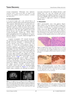
fetoprotein, which lent further support to the possibility of Figure 2. Histopathological images of liver biopsy specimens visualized
hepatobiliary carcinoma. Further imaging with magnetic with hematoxylin and eosin staining. Observations under 20× (A) and
40× (B) magnification show irregularly distributed nests of urothelial
resonance imaging (MRI) showed an 8.5 cm hypoenhancing cells, which are surrounded by fibrotic stroma. Scale bar: (A) 100 µm.
mass within the central aspect of the liver, resulting in (B) 50 µm.
intrahepatic biliary dilatation with multiple other satellite
liver lesions, which were suggestive of cholangiocarcinoma A B
(Klatskin tumor) (Figure 1). Endoscopic retrograde
cholangiopancreatography (ERCP) found a malignant
stricture in the bile duct. Endoscopic ultrasound (EUS)
with negative brush biopsy results led to a liver biopsy to
confirm the diagnosis. Interventional radiology-guided
liver biopsy revealed metastatic carcinoma positive
for GATA3, CK903, P40, P63, and thrombomodulin, Figure 3. Histopathological images of liver biopsy specimens
consistent with metastatic UC (Figures 2-4). The patient visualized with immunohistochemical staining for P40 and high-
denied urinary symptoms, and urinalysis was negative. molecular-weight keratin (HMWK). Observations under 2.5× (A) and
10× (B) magnification show cells positive for P40 and HMWK,
respectively. Scale bar: (A) 1 mm. (B) 500 µm.
A B
A B
Figure 1. T1-weighted axial view image (A) and T2-weighted coronal
view image (B) from the abdominal magnetic resonance imaging scan
show an 8.5 cm hypoenhancing mass within the central aspect of the liver, Figure 4. Histopathological observation of liver specimens visualized
which results in intrahepatic biliary dilatation. Findings are suggestive of with immunohistochemistry at ×10 magnification. The images show
cholangiocarcinoma (Klatskin tumor). Smaller satellite lesions are noted urothelial cells positive for GATA-3. (A) and p63. (B), respectively. Scale
in both lobes of the liver. bar: 500 µm for both panels.
Volume 4 Issue 3 (2025) 97 doi: 10.36922/TD025070011