
Page 109 - TD-4-3
P. 109
Tumor Discovery Mature teratoma during pregnancy
2. Case presentation
A 21-year-old primigravida at a gestational age of 37 weeks
presented with a complaint of sudden abdominal pain
and visited our gynecology department located at the
periphery of Uttar Pradesh, a northern state in India.
Physical examination was normal. Systemic investigations
were carried out, and all were normal. Blood investigation
showed mild leukocytosis. Electrolytes, amylase, lipase,
liver function, and renal function tests were within normal
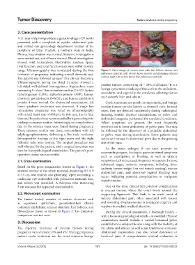
range. Ultrasonography was conducted during the first Figure 1. Gross image of ovarian mass with hair follicle, sebum, and
trimester of pregnancy, indicating a small dermoid cyst. pultaceous material. Left: Ovary shows smooth and glistening external
The patient was followed up upon this clinical discovery. surface; right: Cut section shows hair, pultaceous material.
Ultrasonography during the third trimester showed a 6
left-sided well-defined homogeneous hyperechoic mass ovarian tumors, comprising 24 – 40% of all cases. It is a
measuring 9 × 6 cm. Tumor markers such as CA-125, lactate benign cystic tumor made up of tissues from the endoderm,
dehydrogenase (LDH), alpha-fetoprotein (AFP), human mesoderm, and especially the ectoderm, affecting tissues
7
chorionic gonadotropin (b-hCG), and human epididymis such as teeth, hair, and sebum.
protein 4 were normal. On abdominal examination, left Cystic teratomas are mostly asymptomatic, and benign
lower quadrant tenderness was observed. A single live ovarian tumors are also known as dermoid cysts. In most
intrauterine pregnancy was noted on ultrasonography cases, they are detected incidentally during radiological
with a fetal heart rate of 80 bpm. In this case, due to fetal imaging, routine physical examinations, or pelvic and
distress, the patient was recommended by a gynecologist to abdominal surgeries performed for unrelated conditions.
undergo a cesarean section. Interestingly, no complications When symptoms are present, the most frequently
due to the ovarian mass were found during pregnancy. reported one is lower abdominal or pelvic pain. This may
Then, cesarean section was done, concomitant with left be followed by the discovery of a palpable abdominal
salphingoopherectomy, delivering a live male newborn. or pelvic mass during examination. Some patients may
Intraoperative findings of the right ovary and the right notice an increase in abdominal girth due to the growing
fallopian tube were normal. The surgical procedure was size of the mass.
well tolerated by the patient, and a surgical specimen was As the tumor enlarges, it can exert pressure on
sent for histopathological examination. The patient’s post- surrounding organs, leading to gastrointestinal symptoms
operative course was uneventful.
such as constipation or bloating, as well as urinary
2.1. Gross examination symptoms such as increased frequency or urgency. In more
advanced stages, systemic symptoms, including fever,
Based on the gross examination shown in Figure 1, the cachexia (severe weight loss and muscle wasting), intense
external surface of the ovary received, measuring 9.5 × 9 abdominal pain, and abnormal vaginal bleeding may
× 5.5 cm, was smooth and glistening. Upon sectioning, a occur, indicating potential complications or malignant
unilocular cyst embedded with pultaceous material, hair, transformation.
and sebum was identified. A fallopian tube measuring
3 cm was seen but appeared unremarkable. One of the most critical and common complications
is ovarian torsion, where the ovary twists around the
2.2. Microscopic examination supporting ligaments. This leads to an acute onset of
The tumor mainly consists of mature elements such intense abdominal pain, often associated with nausea
as squamous epithelium, pseudostratified ciliated and vomiting. Ovarian torsion is a surgical exigency and
columnar epithelium, adnexal structure, hair follicles, and requires immediate medical attention.
fibroadipose tissue, as shown in Figure 2. The immature During the clinical assessment, a thorough history –
component was not seen. with a focus on gynecological details – is essential. Physical
3. Discussion examination should include a careful bimanual pelvic
examination to analyze the size along with the mobility of
The reported incidence of ovarian tumors during the uterus and adnexa, as well as any tenderness or masses.
pregnancy varies between 1% and 4%. During pregnancy, Abdominal examination may also reveal distension or
5
mature cystic teratomas are the most common benign localized pain. A comprehensive clinical evaluation is
Volume 4 Issue 3 (2025) 101 doi: 10.36922/TD025120022