
Page 109 - AIH-2-2
P. 109
Artificial Intelligence in Health AI in early breast cancer diagnosis: A review
Traditional diagnostic methods, such as the ones discussed shape, texture, and edge information. Advanced
above, namely mammography, ultrasound, and MRI, have feature extraction methods, such as wavelet
significantly contributed to early detection. However, transforms and texture analysis, are employed to
10
these methods have limitations, including variability in capture intricate details within the images.
interpretation among radiologists, which can lead to missed (iii) Classification: Classification is a fundamental aspect
diagnoses or false positives. CAD systems offer a promising of ML, and these algorithms play a pivotal role in
solution to enhance the accuracy and consistency of early CAD systems. Supervised ML techniques, such as
detection. CAD systems are devised to help radiologists neural networks, support vector machines (SVM),
interpret medical images by highlighting potentially and DL models, are trained on large datasets of
cancerous areas that may require further examination. labeled images to classify regions of interest as either
These systems use complex algorithms combined with benign or malignant. These algorithms learn from
advanced ML techniques to analyze medical images, both seen and unseen data, continuously evolving to
such as mammograms, and identify patterns that indicate improve their accuracy over time.
malignancy. The primary goal of CAD in breast cancer (iv) Detection and localization: The CAD system identifies
detection is to improve diagnostic accuracy, reduce human and localizes suspicious areas within the breast
error, and increase the likelihood of early detection. tissue by marking these regions on the images. This
functionality provides radiologists with visual cues to
2.2.1. Key components of computer-aided detection focus their attention on potential abnormalities. This
systems
step is crucial for ensuring that no suspicious area is
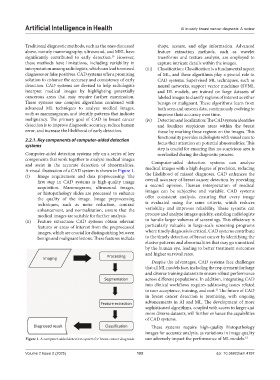
Computer-aided detection systems rely on a series of key overlooked during the diagnostic process.
components that work together to analyze medical images
and assist in the accurate detection of abnormalities. Computer-aided detection systems can analyze
A visual illustration of a CAD system is shown in Figure 1. medical images with a high degree of precision, reducing
(i) Image acquirement and data preprocessing: The the likelihood of missed diagnoses. CAD enhances the
first step in CAD systems is high-quality image overall accuracy of breast cancer detection by providing
acquisition. Mammograms, ultrasound images, a second opinion. Human interpretation of medical
or histopathology slides are processed to enhance images can be subjective and variable. CAD systems
the quality of the image. Image preprocessing offer consistent analysis, ensuring that every image
techniques, such as noise reduction, contrast is evaluated using the same criteria, which reduces
enhancement, and normalization, ensure that the variability and improves reliability. These systems can
medical images are suitable for further analysis. process and analyze images quickly, enabling radiologists
(ii) Feature extraction: CAD systems obtain relevant to handle larger volumes of screenings. This efficiency is
features or areas of interest from the preprocessed particularly valuable in large-scale screening programs
images, which are crucial for distinguishing between where timely diagnosis is critical. CAD systems contribute
benign and malignant lesions. These features include to the timely detection of breast cancer by identifying the
elusive patterns and abnormalities that may go unnoticed
by the human eye, leading to better treatment outcomes
and higher survival rates.
Despite the advantages, CAD systems face challenges
that all ML models face, including the requirement for large
and diverse training datasets to ensure robust performance
across different populations. In addition, integrating CAD
into clinical workflows requires addressing issues related
to user acceptance, training, and cost. The future of CAD
11
in breast cancer detection is promising, with ongoing
advancements in AI and ML. The development of more
sophisticated algorithms, coupled with access to larger and
more diverse datasets, will further enhance the capabilities
of CAD systems.
These systems require high-quality histopathology
images for accurate analysis, as variations in image quality
Figure 1. A computer-aided detection system for breast cancer diagnosis can adversely impact the performance of ML models. 12
Volume 2 Issue 2 (2025) 103 doi: 10.36922/aih.4197