
Page 108 - AIH-2-2
P. 108
Artificial Intelligence in Health AI in early breast cancer diagnosis: A review
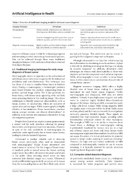
Table 1. Overview of traditional imaging modalities in breast cancer diagnosis
Imaging Technique Advantages Limitations
Mammography Widely available, relatively low cost, effective for Lower sensitivity in dense breast tissue, false positives,
detecting microcalcifications, reduces mortality rates and radiation exposure, requires compression of the
breast
Ultrasound Good for distinguishing solid masses from cysts, no Operator-dependent, lower sensitivity for small tumors,
radiation exposure, useful for dense breast tissue can miss microcalcifications, not effective as a standalone
screening tool
Magnetic resonance imaging Highly sensitive, provides detailed images, useful for Expensive, time-consuming, limited availability, high
high-risk patients, no radiation exposure false-positive rate, may require contrast agents
detection of breast cancer is vital for enhancing prognosis are typically benign. This distinction can be crucial in
and increasing survival rates among breast cancer patients. guiding further diagnostic steps such as biopsies.
This can be achieved through three ways: traditional Although ultrasound is not ideal for initial screening
imaging techniques, CAD, and cancer biomarkers obtained due to limitations in visualizing dense breast tissue, it plays
from liquid biopsies. a vital role in clarifying mammogram findings and aiding
8
2.1. Traditional imaging techniques for early-stage in accurate diagnosis. In addition, ultrasound offers
diagnosis of breast cancer advantages for women with claustrophobia or those with
implants, as it avoids compression and radiation exposure.
Mammography retains its reputation as the cornerstone of While ultrasonography is more sensitive in dense breast
early breast cancer detection, recognized for its widespread tissue, it often misses micro-calcifications characteristic of
availability and cost-effectiveness. This technique uses certain breast cancers. 7
low doses of X-rays to visualize breast tissue in intricate
detail. During a mammogram, a technologist positions Magnetic resonance imaging (MRI) offers a highly
each breast between two paddles, compressing them to detailed view of breast tissue, making it a powerful
achieve optimal image clarity. The X-rays penetrate the instrument for early breast cancer diagnosis. Unlike
breast tissue, with denser areas appearing white and fatty mammography and ultrasound, MRI does not utilize
areas appearing darker in the resulting image. This allows radiation. Instead, it uses high-power magnets and radio
radiologists to identify suspicious abnormalities, such as waves to construct highly detailed three-dimensional
lumps, masses, or calcifications, which are indicative of images of the breasts. During an MRI, a woman lies inside
cancer at its earliest stages. While mammography is highly a large cylindrical scanner while strong magnetic fields
effective, it has limitations. Dense breast tissue, common manipulate water molecules within the body. Radio waves
among younger women, can obscure abnormalities. In are then pulsed, and their interaction with the manipulated
4
addition, some women may experience discomfort during molecules generates detailed signals. These signals are
the compression process. translated into high-resolution images, revealing subtle
abnormalities potentially missed by other techniques.
Despite these limitations, regular mammograms remain The strength of MRI lies in its ability to visualize dense
a crucial tool for early detection, allowing for prompt breast tissue, a challenge for mammography, making
intervention and improved treatment outcomes. However, it particularly beneficial for women who are at a high
in spite of the wide availability of mammography, its risk of breast cancer and those with dense breasts where
sensitivity varies significantly due to breast tissue density, mammograms might miss abnormalities. In addition, MRI
leading to potential missed diagnoses. can assess the extent of a tumor and analyze its potential
Ultrasound is another valuable tool in detecting spread to lymph nodes, assisting in treatment planning.
9
breast cancer, particularly when used in conjunction However, the high cost and limited availability of MRI
with mammography. Unlike X-rays, ultrasound uses compared to mammography restricts its use in routine
4
sound waves to construct life-like images of breast tissue. screening. It is typically reserved for specific situations
A handheld probe is moved across the breast, emitting requiring clearer diagnostic insights.
sound waves that reflect off internal structures and return
to the probe. These returning sound waves are then 2.2. Computer-aided detection/diagnosis
converted into detailed images on a screen. Ultrasound As previously established, breast cancer is a widespread
excels at differentiating potentially cancerous solid masses cancer among women worldwide. Early detection
from the surrounding tissues and fluid-filled cysts, which is warranted for better prognosis and survival rates.
Volume 2 Issue 2 (2025) 102 doi: 10.36922/aih.4197