
Page 52 - AN-1-1
P. 52
Advanced Neurology EPAC2 null leads to tauopathy
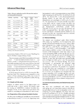
Table 1. Primary antibodies used for Western blot analysis homogenized in radio immunoprecipitation assay (RIPA)
and immunohistochemistry buffer containing 50 mM Tris-HCl, 150 mM NaCl, 20 mM
EDTA, 1% Nonidet-P40, 1 mM phenylmethylsulfonyl
Antibody Specificity Type Dilution Dilution Source
(WB) (IHC) fluoride (PMSF), 50 mM NaF, and 0.25% sodium
pT205 P-tau at Thr205 pAb 1:1000 1:200 Biosource deoxycholate, and centrifuged at 12,000 ×g for 30 min at
4°C. The supernatant was collected, and after evaporation
pT231 P-tau at Ser231 pAb 1:1000 Biosource of the formic acid in a speed vacuum, the pellet was
pS396 P-tau at Ser396 pAb 1:1500 1:200 Biosource resuspended in sodium dodecyl sulfate-polyacrylamide
pS404 P-tau at Ser404 pAb 1:1500 Biosource gel electrophoresis (SDS-PAGE) sample buffer
P35/25 Total P25/25 pAb 1:1000 Cell (240 mM Tris-HCl, pH 6.8, 6% SDS, 30% glycerol, and
signaling 0.06% bromophenol blue). The RIPA fraction and the
CDK5 Total CDK5 mAb 1:1000 Abcam formic acid fraction contained relatively soluble tau and
P35 P35 pAb 1:500 Santa Crus detergent-insoluble tau, respectively, and were sonicated
p35/p25 P35 and P25 mAb 1:500 Cell extensively before immunoblotting.
signaling 2.7. Western blotting
TAU-1 Non- mAb 1:500 Millipore
phosphorylated The animals were decapitated under chloral hydrate
tau (600 mg/kg, i.p.) anesthesia, and the hippocampi were
TAU-46 Total tau mAb 1:1000 Millipore rapidly removed and homogenized at 4°C using a Teflon
EPAC2 Epac2 pAb 1:500 Santa Crus glass homogenizer in a reagent containing 50 mM Tris-
DM1A α-tubulin mAb 1:1000 Sigma HCl (pH 7.4), 1 mM Na VO , 150 mM NaCl, 10 mM
3
4
NaF, 5 mM EDTA, 2 mM benzamidine, 1 mM PMSF,
WB: Western blot; IHC: Immunohistochemistry; pAb: Polyclonal and proteinase inhibitor cocktail (1:100 dilution). Three
antibody; mAb: Monoclonal antibody.
volumes of the homogenized tissue were added to one
reverse transcribed to cDNA with the use of FastKing One volume of extraction buffer containing 200 mM Tris-HCl,
Step RT-PCR kit (Tiangen, Beijing, China). pH 7.6, 8% SDS, 40% glycerol, and 40 mM dithiothreitol,
and then, the mixture was placed in a boiling water
The primer sequences for EPAC2 were as follows: (Forward bath for 10 min. Next, the lysates were sonicated briefly
primer) 5ʹ-AACTGGTATGCTGTCCTGGC-3ʹ and and centrifuged at 12,000 ×g for 5 min. The supernatant
(reverse primer) 5ʹ-TAGGGAGGAGCCAGAAGTCC-3ʹ. was stored at −80°C for Western blotting. The protein
The primer sequences for β-actin were as follows: (Forward concentration of supernatants was measured using
primer) 5ʹ-AGCCTTCCTTCTTGGGTAT-3ʹ and (reverse the Pierce BCA Protein Assay Kit, according to the
primer) 5ʹ-GCTCAGTAACAGTCCGCCTA-3ʹ. The manufacturer’s instructions. The proteins were separated
primers used in this experiment were produced by Tsingke by 10% SDS-PAGE and transferred to a nitrocellulose
Biotechnology (Beijing, China). membrane. After blocking in 3% bovine serum albumin
for 1 h at 25°C, the membranes were incubated first with
RT-PCR was performed on a PCR thermocycler (Bio- primary antibodies at 4°C overnight and then with anti-
Rad, New York, USA). Reactions were prepared in a total mouse or anti-rabbit IgG conjugated to IRDye 800CW as
volume of 50 μL containing 0.5 μg cDNA, 2 μL of each 10 secondary antibody for 1 h at 25°C. Protein expression was
μM primer, and 25 μL of Taq MasterMix (Cwbio, Beijing, then visualized with the Odyssey Infrared Imaging System
China). (LI-COR Biosciences).
2.5. Calpain activity assay 2.8. Immunohistochemistry and Bielschowsky silver
Analysis of calpain activity in the hippocampus was staining
performed using a calpain activity fluorometric assay The mice were anesthetized with chloral hydrate
kit (catalog no. GMS50046.2, Genmed Scientifics Inc., (600 mg/kg, i.p.) and immediately perfused with 200 mL
Shanghai, China). All the experimental procedures were normal saline followed by 200 ml phosphate buffer (4°C)
carried out, according to the manufacturer’s instructions. containing 4% paraformaldehyde. The brains were then
post-fixed in perfusate at 4°C for 24 h. Paraffin-embedded
2.6. Preparation of the sarkosyl-insoluble fraction
brain sections were prepared for immunostaining after
The detection of soluble and insoluble tau was performed xylene treatment and progressive rehydration with 70–100%
as described previously . In brief, the hippocampi were ethanol. Sections were blocked and then incubated with
[20]
Volume 1 Issue 1 (2022) 3 https://doi.org/10.36922/an.v1i1.8