
Page 55 - AN-1-1
P. 55
Advanced Neurology EPAC2 null leads to tauopathy
A B C
D
E F
G
H
I
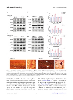
Figure 3. Abnormal tau aggregation and neuronal morphology in EPAC2 mice. (A–H) Hippocampus extracts from 6-month-old and 18-month-
−/−
old EPAC2 mice and wild-type EPAC2 littermates. (A and B) Representative blot images for soluble tau. (E and F) Representative blot images for
−/−
+/+
insoluble tau. The densitometric analyses of the immunoreactivities to the antibodies indicated in (A and B) and (E and F) are shown in (C and D) and
(G and H), respectively; n = 3; *P < 0.05; **P < 0.01. (I) Cortical slices from 9-month-old EPAC2 mice and wild-type EPAC2 littermates were subjected
−/−
+/+
to Bielschowsky silver staining. Representative images with magnifications are shown.
GSK-3β, JNK, and CDK5, all of which are critical regulators JNK (t-JNK), or phosphorylated Thr183/185 in JNK
of tau hyperphosphorylation. We found that the absence of (p-JNK) (Figure 4A and B). However, CDK5 levels and
−/−
EPAC2 did not induce any changes in the protein levels of the p25/p35 ratio were dramatically increased in EPAC2
the catalytic subunit of PP2a (PP2ac), β subunit of PP2a mice (Figure 4C and D), suggesting that CDK5 activation
(PP2ab), total ERK (t-ERK), phosphorylated Thr202/ might be involved in EPAC2 loss-induced tau pathology.
Tyr204 in ERK (p-ERK), total GSK-3β (t-GSK-3β), Consistent with these observations, silencing of EPAC2
phosphorylated serine 9 in GSK-3β (pS9-GSK-3β), total in the N2a cells also resulted in the activation of CDK5,
Volume 1 Issue 1 (2022) 6 https://doi.org/10.36922/an.v1i1.8