
Page 101 - AN-3-4
P. 101
Advanced Neurology Shuntogram technique in programmable valves
to the valve. This presentation, however, may also occur position for 5 min, and the scan was repeated, yet no flow
in patent shunts. Complete occlusion of the peritoneal was detected. Examination of the shunt valve revealed
catheter results in no distal flow, though tracer may still that it had inadvertently been set to 5 – a relatively high-
appear in the ventricular system, spinal canal, and kidneys. pressure setting for this device. To confirm that this high
Partial occlusions are identified by slow tracer transit or setting was causing the flow failure, and the shunt was
accumulation, while a disconnection often results in adjusted to the lowest possible setting. Upon re-imaging,
tracer widening or accumulating at the discontinuity site. immediate counts were noted in the distal tubing and
Occlusion involving the valve itself may produce symptoms abdomen (Figure 1B and C). At the conclusion of the
resembling proximal or distal obstruction, depending on procedure, the shunt setting was re-adjusted to 4, with
the valve’s position relative to the reservoir. 25,26 plans for close follow-up in the outpatient clinic. Following
this intervention, the patient experienced improvement
2.1. Case presentation
without the need for surgery. The radiologist remarked that
2.1.1. The case he would have initially diagnosed the shunt as occluded
Here, we describe a case of a 73-year-old male diagnosed based on the initial images and acknowledged that he had
with normal pressure hydrocephalus who had previously gained valuable insight from this case.
undergone placement of an adjustable right occipital 2.1.2. Shunt protocol
ventriculoperitoneal shunt (Codman Certas, Integra
LifeSciences, New Jersey). The patient presented to The patient is positioned in a semirecumbent posture,
our neurosurgery clinic with intermittent episodes of and the site is prepped without shaving, using at least
cognitive decline and gait/balance issues despite stable two chlorhexidine prep sticks (ChloraPrep™, Becton,
imaging. These symptoms prompted an evaluation of Dickinson in Franklin Lakes, NJ, USA) and some
shunt patency through a shunt function study. It was abrasive cleansing of the site. This seated positioning
unclear from his records if the shunt valve pressure had is preferred during preparation to avoid temporarily
been adjusted during previous clinic visits. The patient disturbing the shunt system’s fluid dynamics, although
was referred to the Department of Nuclear Medicine for a patients who are bed-confined may remain lying down.
shuntogram, and coordination of contrast administration Using a sterile technique, a 25-gauge butterfly needle is
was performed by a neurosurgery resident. The patient inserted to access the shunt reservoir. Correct needle
underwent standard preparation with chlorhexidine and placement is confirmed by withdrawing a small volume
alcohol, and shaving was not performed. Under sterile of CSF, typically < 0.1 mL. A tuberculin (1 mL) syringe
technique, the right occipital shunt reservoir was accessed is then used to inject a volume of radiotracer containing
with a 25-gauge butterfly needle, and a 400 μL solution technetium-99, usually between 0.1 and 0.2 mL, strictly no
containing technetium-99 was injected into the reservoir. more than 0.5 mL. Care must be taken to avoid significant
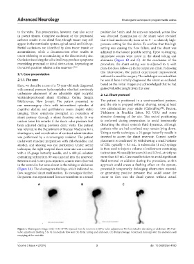
Between 0 and 5-min post-injection, counts were observed fluid removal or addition during the procedure, as this
in the ventricles but were absent in the tubing or abdomen approach could create a flushing effect on the system,
(Figure 1A). The shuntogram findings, which indicated no potentially temporarily dislodging obstructive material
flow, suggested shunt malfunction. To investigate further, or generating positive pressure that could cause the
the patient was repositioned from a recumbent to a seated tracer to flow into the distal system without actual
A B C
Figure 1. Shuntogram images with 111In-DTPA injected into the reservoir. (A) Pre-valve adjustment: No flow noted in the tubing or abdomen. (B) Post-
valve adjustment (Setting 5 to 1): Immediate flow into the distal tubing and abdomen. (C) Delayed image: Continued drainage into the abdomen and
emptying of the ventricle
Volume 3 Issue 4 (2024) 3 doi: 10.36922/an.4180