
Page 80 - AN-3-4
P. 80
Advanced Neurology SARS-CoV-2 in age-associated neurodegeneration
where viral antigen was detected in the olfactory bulb immune response, which ensures its persistence over an
3 days after intranasal inoculation in mice. The virus then extended period (Table 2).
spreads to the cortex, mesolimbic cortex, hippocampus,
amygdala, and finally to the brainstem and spinal cord 5. The role of SARS-CoV-2 proteins in viral
within 7 days. Ablation of the olfactory bulb after nasal persistence in the brain by manipulating
infection with MHV blocked further spread, supporting immunoregulatory pathways
the theory of spread through the olfactory tract. 63
Numerous clinical studies on long COVID have shown
An alternative route of transmission is through the that viral particles are present in the brain and affect
vagus nerve and the GI tract, which may play a central brain architecture. 5,17,40 However, it remains crucial
role in the retrograde penetration of SARS-CoV-2 into the to understand how SARS-CoV-2 manipulates host
CNS. Another important mechanism is the extracellular immunoregulatory mechanisms, contributing to persistent
64
vesicular transport of SARS-CoV-2 or key components neuroinflammation (Figure 3). The most important
of its proteome from the site of primary infection to the strategy of SARS-CoV-2, shared by other coronaviruses, is
CNS. Neuronally enriched extracellular vesicles, including replicating within double-membrane vesicles. This strategy
exosomes from individuals with PASC, are enriched with prevents the activation of retinoic acid-inducible gene
markers of neurodegeneration, such as amyloid, low- 1 (RIG-I)-like receptors, which recognize viral double-
molecular-weight neurofilament subunit protein, total tau, stranded RNA intermediates. 68,69
phosphorylated tau, and neurogranin. This enrichment
suggests that these vesicles may play a critical role in SARS-CoV-2 is not only adept at evading detection
70
the amplification of AD pathology in patients following but also at disguising itself. Chen et al. showed that
COVID infection. 65,66 In addition, membrane-bound the non-structural protein (Nsp) 14 of SARS-CoV-2
exosomes originating from the lungs, which contain possesses guanine N7 methyltransferase activity, which
transcription factors linked to neuronal gene regulation allows it to mimic the cap structure on viral RNA. In
in Alzheimer’s and Parkinson’s diseases, have been addition, the Nsp16 protein of SARS-CoV-2 modifies
documented to be transported into the brain through the this cap-like structure through its 2’O-methyltransferase
trans-neuronal pathway. Cumulatively, SARS-CoV-2 activity, enabling the virus to evade recognition by
67
utilizes multiple mechanisms to disable and evade the host melanoma differentiation-associated protein-5 (MDA5).
71
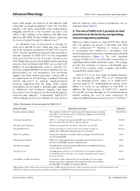
Table 2. Mechanisms of neuroinvasion by SARS‑CoV‑2
Mode of invasion Mechanism of invasion References
Receptor-mediated pathway The s-protein of SARS-CoV-2 binds to the ACE2 receptor and co-receptor 44,46,47,50,51
neuropilin-1; host proteases such as TMPRSS2, cathepsin L, and furin help
in the cleavage of the s-protein
Hematogenous pathway SARS-CoV-2 disrupts the alveolar epithelial barrier following primary 45,46,48
infection and reaches the CNS through bloodstream
Through blood-CSF barrier The infection spreads from the blood–CSF barrier into the CNS 46,54
Through BBB barrier Heightened interferon response and cytokine storm may alter the 46,52,55
architecture of BBB, thus making it permeable to the virus
Trojan horse pathway The infected leukocytes, monocytes, and macrophages may infiltrate into 46,56
the CNS through BBB
Trans neuronal pathway SARS-CoV-2 may initially infect the peripheral nerve endings and then 46,58
enter the CNS through a synapse-connected route
The olfactory pathway The viral particles reach the olfactory bulb via the nasal-epithelial pathway 46,60
and then spread into the CNS
Through GI tract The spike-protein of SARS-CoV-2 binds to an ACE2 receptor present on 46,62
the epithelial cells lining the gut, and through retrograde axonal transport,
it reaches the CNS
Extracellular vesicular transmission Extracellular vesicles released from alveolar epithelial cells may contain 46,63,65
viral genome or key components of viral proteome that reach to CNS by
invading BBB or through trans-neuronal pathway
Abbreviations: ACE2: Angiotensin-converting enzyme 2; BBB: Blood–brain barrier; CNS: Central nervous system; CSF: Cerebrospinal fluid;
SARS-CoV-2: Severe acute respiratory syndrome-coronavirus-2; TMPRSS2: Transmembrane protease serine 2.
Volume 3 Issue 4 (2024) 7 doi: 10.36922/an.4267