
Page 31 - BH-2-1
P. 31
Brain & Heart Pictorial rendition pulmonary stenosis
A B
Figure 49. Cineangiographic images of the pulmonary artery immediately
before (A) and 12 months after (B) balloon pulmonary valvuloplasty
(BPV) in an infant with Fallot’s tetralogy with subvalvar and valvar
pulmonary obstruction, illustrating improved diameter of the main
Figure 47. The outcome of balloon pulmonary valvuloplasty (BPV) on pulmonary artery (MPA) at follow-up. The left pulmonary artery (LPA)
blood flow to the lungs (Qp) in l/min/m (left group) and the ratio of blood and right pulmonary artery (RPA) are labeled. Replicated from Rao et al. 78
2
flow between pulmonary and systemic circuits (Qp: Qs) (right group)
both at the time of BPV and at follow-up (FU). A significant (P < 0.01) A B
increase in Qp and Qp: Qs occurred after BPV. These measurements
remained unaltered (P > 0.1) at FU. However, the standard errors of mean
(SEM) are larger at FU. Replicated from Rao. 48
A B
C D
Figure 48. Cineangiographic images of pulmonary arteries immediately
before (A) and 6 months following (B) balloon pulmonary valvuloplasty
(BPV) in an infant with transposed great vessels, ventricular septal defect,
and subvalvar and valvar pulmonary obstruction, illustrating improved
diameters of the pulmonary arteries both on the right (RPA) and left
(LPA) sides. There is a variation in magnification; both cineangiograms
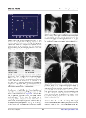
were performed with catheters of #5-F size. Following adjustment for Figure 50. Balloon dilatation catheters positioned across the porcine
magnification, the RPA improved from 5.0 mm to 9.4 mm and the LPA heterografts (Hancock). The partially inflated balloon (A) shows the
from 3.3 mm to 7.8 mm. Replicated from Rao and Brais. 77 balloon “waisting” (arrowheads), almost entirely eliminated (B) with
further balloon inflation. Fully inflated balloons are featured in two other
the pulmonary valve is higher than 50 mmHg, dilation of patients (C and D). It is observed that waisting (arrows) persists to some
such valve becomes necessary. The procedure of BPV extent in these two frames. In addition, note that the waisting (arrows)
19
mirrors that used for native PS (Figure 50). It is imperative is just distal to the opaque metallic ring of the Hancock prosthesis. In
(D), the connection between the right ventricle and conduit is marked
that the balloon’s diameter matches that of the initially by another ring (R). The pigtail (PT) catheter in the ascending aorta is
implanted valve. 19,87 In the study, the patients had an labeled. Reproduced from Rao. 19
average age of 14.9 ± 6.2 years at the time of BPV. For the
19
entire cohort, the peak systolic pressure difference through decreased from 108 ± 33 to 88 ± 24 mmHg, and the right
the porcine heterograft reduced from 77 ± 25 to 46 ± ventricle/left ventricle peak systolic pressure ratio declined
29 mmHg, the peak systolic pressure in the right ventricle from 0.94 ± 0.19 to 0.72 ± 0.22. While these results may
Volume 2 Issue 1 (2024) 15 https://doi.org/10.36922/bh.2406