
Page 30 - BH-2-1
P. 30
Brain & Heart Pictorial rendition pulmonary stenosis
A B A
Figure 43. The balloon pulmonary valvuloplasty (BPV) procedure B
entails the insertion of a balloon valvuloplasty catheter through the
stenotic pulmonary valve, followed by inflation with diluted contrast
material. Balloon waisting is observed (A) as the balloon inflated
(arrows). This waisting is caused by the narrowed pulmonary valve and
disappears (arrow in [B]) with further balloon inflation, resulting in the
relief of pulmonary valve obstruction. Only lateral views are displayed.
Reproduced from Rao et al. 78
A B
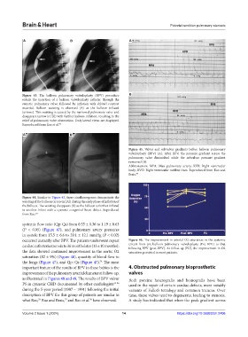
Figure 45. Valvar and subvalvar gradients before balloon pulmonary
valvuloplasty (BPV) (A). After BPV, the pressure gradient across the
pulmonary valve diminished while the subvalvar pressure gradient
remained (B).
Abbreviations: MPA: Man pulmonary artery; RVB: Right ventricular
body; RVO: Right ventricular outflow tract. Reproduced from Rao and
Brais. 77
Figure 44. Similar to Figure 43, these cinefluorograms demonstrate the
waisting of the balloon (arrow in [A]) during the early phase of inflation of
the balloon. The waisting disappears (B) as the balloon is further inflated
in another infant with a cyanotic congenital heart defect. Reproduced
from Rao. 20
systemic flow ratio (Qp: Qs) from 0.55 ± 0.36 to 1.19 ± 0.63
(P < 0.05) (Figure 47), and pulmonary artery pressures
in systole from 15.5 ± 6.6 to 29.1 ± 12.1 mmHg (P < 0.02)
occurred instantly after BPV. The patients underwent repeat Figure 46. The improvement in arterial O2 saturation in the systemic
cardiac catheterization six to 36 months later (13 ± 10 months); circuit from pre-balloon pulmonary valvuloplasty (Pre-BPV) to that
following BPV (post-BPV). At follow-up (FU), the improvement in O2
the data showed continued improvement in the aortic O2 saturation persisted in most patients.
saturation (82 ± 9%) (Figure 46), quantity of blood flow to
the lungs (Figure 47), and Qp: Qs (Figure 47). The most
78
important feature of the results of BPV in these babies is the 4. Obstructed pulmonary bioprosthetic
improvement of the pulmonary arterial diameter at follow-up, valves
as illustrated in Figures 48 and 49. The results of BPV valvar Both porcine heterografts and homografts have been
PS in cyanotic CHD documented by other cardiologists 79-86 used in the repair of certain cardiac defects, most notably
during the 5-year period (1987 – 1991) following the initial variants of Fallot’s tetralogy and common truncus. Over
description of BPV for this group of patients are similar to time, these valves tend to degenerate, leading to stenosis.
what Rao, Rao and Brais, and Rao et al. have observed. A study has indicated that when the peak gradient across
76
77
78
Volume 2 Issue 1 (2024) 14 https://doi.org/10.36922/bh.2406